The Phospho-STAT1-S727 Monoclonal Antibody (CABP1000) is a high-quality antibody developed for reliable detection and analysis of target proteins. This antibody, developed by Assay Genie, is specifically designed for use in various applications such as Western blot and immunohistochemistry to detect and analyze the phosphorylation status of STAT1 at serine 727 in human samples.STAT1 is a key transcription factor involved in the regulation of immune response, cell growth, and differentiation. Phosphorylation of STAT1 at serine 727 is known to enhance its transcriptional activity and influence its function in various cellular processes.
This antibody is validated for use in WB, IHC-P, IF/ICC, ELISA applications and has demonstrated reactivity against Human, Mouse, Rat samples.
Product Name:
Phospho-STAT1-S727 Monoclonal Antibody
SKU:
CABP1000
Size:
20μL, 100μL
Reactivity:
Human, Mouse, Rat
Clone Number:
ARC1544
Conjugate:
Unconjugated
Immunogen:
Synthetic peptide. This information is considered to be commercially sensitive.
Sequence:
PMSP E
Tested Applications:
WBIHC-PIF/ICCELISA
Recommended Dilution:
WB
1:13000 - 1:26000
IF/ICC
1:1000 - 1:4000
IHC-P
1:300 - 1:1200
ELISA
Recommended starting concentration is 1 μg/mL. Please optimize the concentration based on your specific assay requirements.
The protein encoded by this gene is a member of the STAT protein family. In response to cytokines and growth factors, STAT family members are phosphorylated by the receptor associated kinases, and then form homo- or heterodimers that translocate to the cell nucleus where they act as transcription activators. The protein encoded by this gene can be activated by various ligands including interferon-alpha, interferon-gamma, EGF, PDGF and IL6. This protein mediates the expression of a variety of genes, which is thought to be important for cell viability in response to different cell stimuli and pathogens. The protein plays an important role in immune responses to viral, fungal and mycobacterial pathogens. Mutations in this gene are associated with Immunodeficiency 31B, 31A, and 31C.
Purification Method
Affinity purification
Gene ID
6772
RRID
AB_2863892
Buffer Information
Store at -20℃. Avoid freeze / thaw cycles. Buffer: PBS containing 50% glycerol and 0.05% BSA, preserved with proclin300 or sodium azide, pH 7.3.
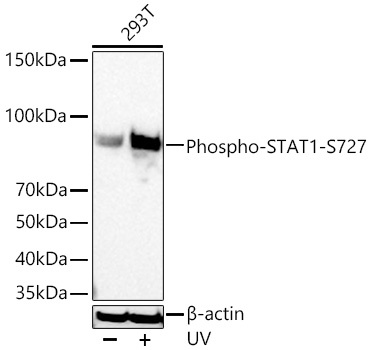
Western blot analysis of lysates from 293T cells using Phospho-STAT1-S727 Rabbit mAb (CABP1000) at 1:13000 dilution incubated overnight at 4℃. 293T cells were treated with UV at room temperature for 15-30 minutes. Secondary antibody: HRP-conjugated Goat anti-Rabbit IgG (H+L) (CABS014) at 1:10000 dilution. Lysates/proteins: 30 μg per lane. Blocking buffer: 3 % nonfat dry milk in TBST. Detection: ECL Basic Kit (AbGn00020). Exposure time: 5s.
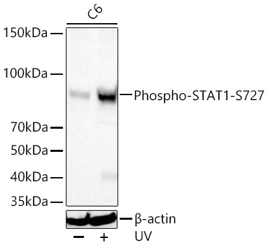
Western blot analysis of lysates from C6 cells using Phospho-STAT1-S727 Rabbit mAb (CABP1000) at 1:13000 dilution incubated overnight at 4℃. C6 cells were treated with UV at room temperature for 15-30 minutes. Secondary antibody: HRP-conjugated Goat anti-Rabbit IgG (H+L) (CABS014) at 1:10000 dilution. Lysates/proteins: 30 μg per lane. Blocking buffer: 3 % nonfat dry milk in TBST. Detection: ECL Basic Kit (AbGn00020). Exposure time: 45s.
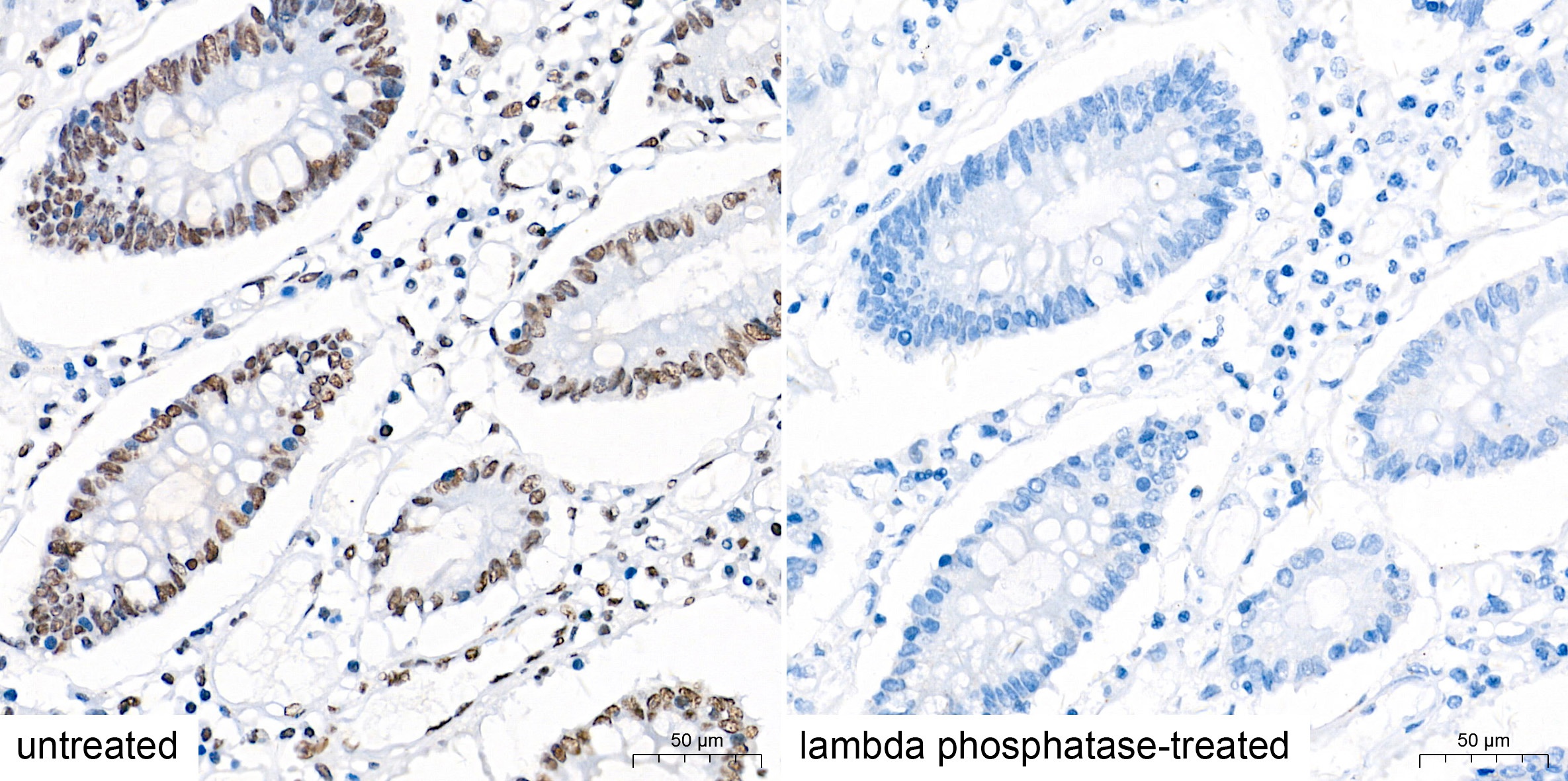
Immunohistochemistry analysis of paraffin-embedded Human colon tissue using Phospho-STAT1-S727 Rabbit mAb (CABP1000) at a dilution of 1:500 (40x lens). High pressure antigen retrieval performed with 0.01M Tris-EDTA Buffer (pH 9.0) prior to IHC staining.
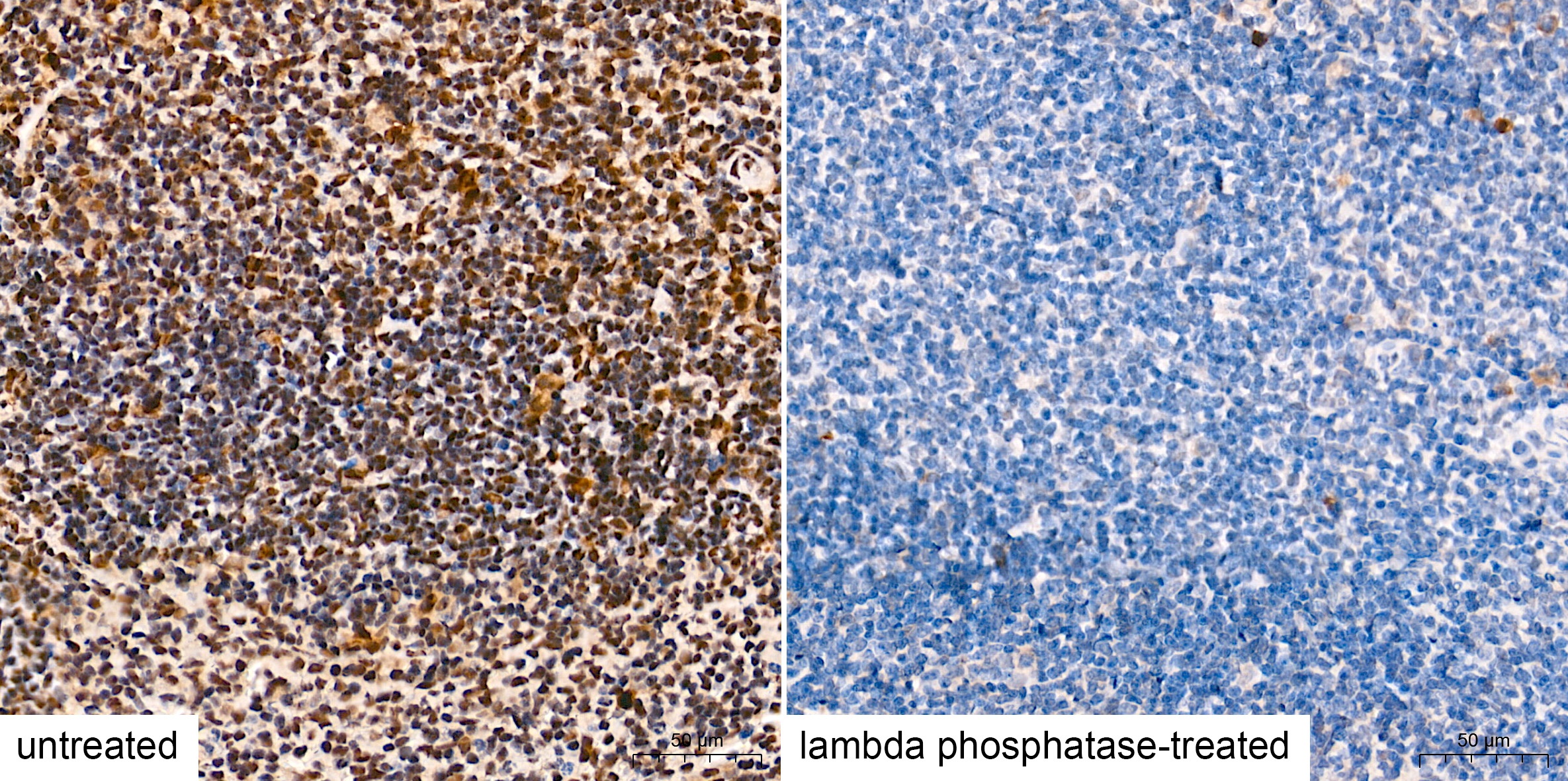
Immunohistochemistry analysis of paraffin-embedded Mouse spleen tissue using Phospho-STAT1-S727 Rabbit mAb (CABP1000) at a dilution of 1:500 (40x lens). High pressure antigen retrieval performed with 0.01M Tris-EDTA Buffer (pH 9.0) prior to IHC staining.
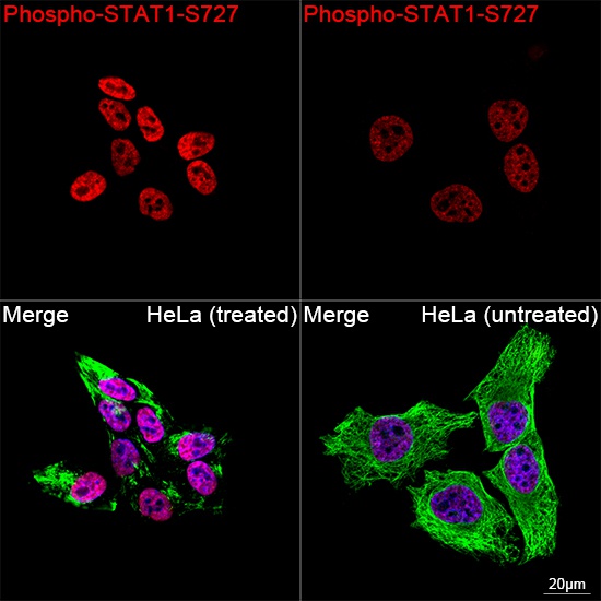
Confocal imaging of HeLa cells (treated with hIFN-α1) and HeLa cells (untreated) cells using Phospho-STAT1-S727 Rabbit mAb (CABP1000, dilution 1:2000) followed by a further incubation with Cy3 Goat Anti-Rabbit IgG (H+L) (CABS007, dilution 1:500) (Red). The cells were counterstained with α-Tubulin Mouse mAb (AC012, dilution 1:400) followed by incubation with ABflo® 488-conjugated Goat Anti-Mouse IgG (H+L) Ab (CABS076, dilution 1:500) (Green). DAPI was used for nuclear staining (Blue). Objective: 100x.
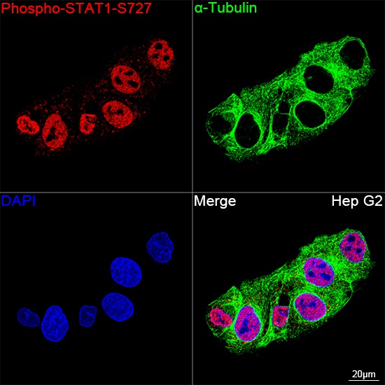
Confocal imaging of Hep G2 cells using Phospho-STAT1-S727 Rabbit mAb (CABP1000, dilution 1:2000) followed by a further incubation with Cy3 Goat Anti-Rabbit IgG (H+L) (CABS007, dilution 1:500) (Red). The cells were counterstained with α-Tubulin Mouse mAb (AC012, dilution 1:400) followed by incubation with ABflo® 488-conjugated Goat Anti-Mouse IgG (H+L) Ab (CABS076, dilution 1:500) (Green). DAPI was used for nuclear staining (Blue). Objective: 100x.
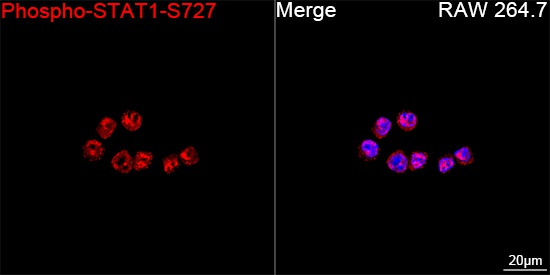
Confocal imaging of RAW 264.7 cells using Phospho-STAT1-S727 Rabbit mAb (CABP1000, dilution 1:2000) followed by a further incubation with Cy3 Goat Anti-Rabbit IgG (H+L) (CABS007, dilution 1:500) (Red). DAPI was used for nuclear staining (Blue). Objective: 100x.
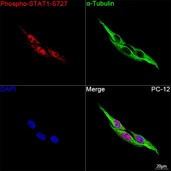
Confocal imaging of PC-12 cells using Phospho-STAT1-S727 Rabbit mAb (CABP1000, dilution 1:2000) followed by a further incubation with Cy3 Goat Anti-Rabbit IgG (H+L) (CABS007, dilution 1:500) (Red). The cells were counterstained with α-Tubulin Mouse mAb (AC012, dilution 1:400) followed by incubation with ABflo® 488-conjugated Goat Anti-Mouse IgG (H+L) Ab (CABS076, dilution 1:500) (Green). DAPI was used for nuclear staining (Blue). Objective: 100x.