The PPIL4 Antibody (CAB15920) is a high-quality antibody developed for reliable detection and analysis of target proteins. This antibody, produced in rabbits, exhibits high reactivity with human samples and has been validated for use in Western blot applications. By specifically binding to the PPIL4 protein, this antibody enables detection and analysis in a variety of cell types, making it an ideal choice for investigations in molecular biology and cell signaling research.PPIL4, also known as a member of the cyclophilin family, is involved in protein folding and maturation, making it a key player in maintaining cellular homeostasis. Its role in regulating protein function and stability highlights its importance in processes like cell growth, differentiation, and apoptosis.
This antibody is validated for use in WB, IF/ICC, ELISA applications and has demonstrated reactivity against Human samples.
Product Name:
PPIL4 Antibody
SKU:
CAB15920
Size:
20μL, 100μL
Reactivity:
Human
Conjugate:
Unconjugated
Immunogen:
Recombinant protein (or fragment).This information is considered to be commercially sensitive.
Recommended starting concentration is 1 μg/mL. Please optimize the concentration based on your specific assay requirements.
Synonyms:
HDCME13P, PPIL4
Positive Sample:
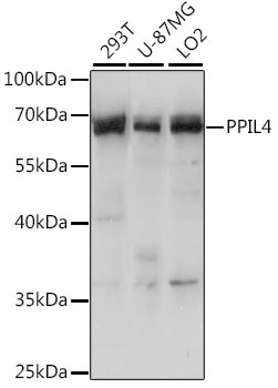
293T, U-87MG, LO2
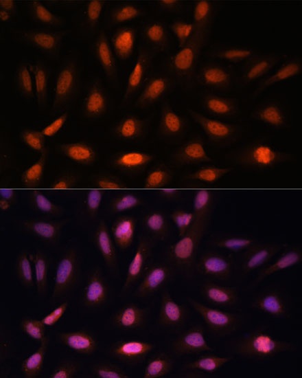
Cellular Localization:
Nucleus.
Calculated MW:
57kDa
Observed MW:
65kDa
This gene is a member of the cyclophilin family of peptidylprolyl isomerases. The cyclophilins are a highly conserved family, members of which play an important role in protein folding, immunosuppression by cyclosporin A, and infection of HIV-1 virions.
Purification Method
Affinity purification
Gene ID
85313
RRID
AB_2763353
Buffer Information
Store at -20℃. Avoid freeze / thaw cycles. Buffer: PBS with 0.01% thimerosal,50% glycerol,pH7.3.
Western blot analysis of various lysates using PPIL4 Rabbit pAb (CAB15920) at 1:1000 dilution. Secondary antibody: HRP-conjugated Goat anti-Rabbit IgG (H+L) (CABS014) at 1:10000 dilution. Lysates/proteins: 25μg per lane. Blocking buffer: 3% nonfat dry milk in TBST. Detection: ECL Basic Kit (AbGn00020). Exposure time: 10s.
Immunofluorescence analysis of U-2 OS cells using PPIL4 Rabbit pAb (CAB15920) at dilution of 1:100. Secondary antibody: Cy3-conjugated Goat anti-Rabbit IgG (H+L) (CABS007) at 1:500 dilution. Blue: DAPI for nuclear staining.