The RPS12 Antibody (CAB5890) is a high-quality antibody developed for reliable detection and analysis of target proteins. This antibody, raised in rabbits, exhibits high reactivity with human samples and is validated for use in Western blot applications. By binding to the RPS12 protein, researchers can detect and analyze its expression in various cell types, making it ideal for studies in molecular biology and protein synthesis research.RPS12 is essential for ribosomal function, playing a key role in the translation of genetic information into functional proteins. Dysregulation of RPS12 has been linked to various diseases, including cancer and genetic disorders.
This antibody is validated for use in WB, IF/ICC, ELISA applications and has demonstrated reactivity against Human, Mouse, Rat samples.
Product Name:
RPS12 Antibody
SKU:
CAB5890
Size:
20μL, 100μL
Reactivity:
Human, Mouse, Rat
Conjugate:
Unconjugated
Immunogen:
Recombinant protein (or fragment).This information is considered to be commercially sensitive.
Ribosomes, the organelles that catalyze protein synthesis, consist of a small 40S subunit and a large 60S subunit. Together these subunits are composed of 4 RNA species and approximately 80 structurally distinct proteins. This gene encodes a ribosomal protein that is a component of the 40S subunit. The protein belongs to the S12E family of ribosomal proteins. It is located in the cytoplasm. Increased expression of this gene in colorectal cancers compared to matched normal colonic mucosa has been observed. As is typical for genes encoding ribosomal proteins, there are multiple processed pseudogenes of this gene dispersed through the genome.
Purification Method
Affinity purification
Gene ID
6206
RRID
AB_2766638
Buffer Information
Store at -20℃. Avoid freeze / thaw cycles. Buffer: PBS containing 50% glycerol, preserved with proclin300 or sodium azide, pH 7.3.
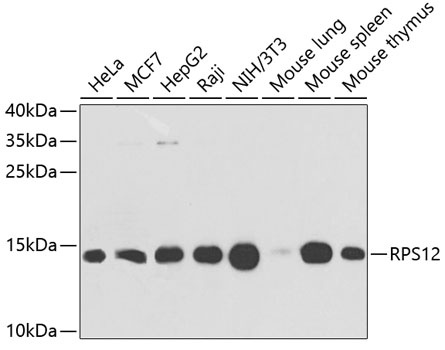
Western blot analysis of various lysates using RPS12 Rabbit pAb (CAB5890) at 1:1000 dilution. Secondary antibody: HRP-conjugated Goat anti-Rabbit IgG (H+L) (CABS014) at 1:10000 dilution. Lysates/proteins: 25μg per lane. Blocking buffer: 3% nonfat dry milk in TBST. Detection: ECL Basic Kit (AbGn00020). Exposure time: 15s.
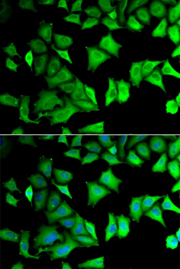
Immunofluorescence analysis of MCF-7 cells using RPS12 Rabbit pAb (CAB5890). Secondary antibody: Cy3-conjugated Goat anti-Rabbit IgG (H+L) (CABS007) at 1:500 dilution. Blue: DAPI for nuclear staining.