The SCO1 Antibody (CAB6734) is a high-quality antibody developed for reliable detection and analysis of target proteins. This antibody, produced in rabbits, is highly specific to human samples and has been validated for use in Western blot applications. By binding to the SCO1 protein, this antibody allows for the detection and analysis of SCO1 levels in various cell types, making it ideal for investigations in the fields of biochemistry and cell biology.SCO1 is a critical component of the mitochondrial respiratory chain, playing a key role in cellular energy production. Dysregulation of SCO1 has been linked to various diseases, including mitochondrial disorders and neurodegenerative conditions.
This antibody is validated for use in WB, IHC-P, ELISA applications and has demonstrated reactivity against Human, Mouse, Rat samples.
Product Name:
SCO1 Antibody
SKU:
CAB6734
Size:
20μL, 100μL
Reactivity:
Human, Mouse, Rat
Conjugate:
Unconjugated
Immunogen:
Recombinant protein (or fragment).This information is considered to be commercially sensitive.
Recommended starting concentration is 1 μg/mL. Please optimize the concentration based on your specific assay requirements.
Synonyms:
SCOD1, MC4DN4, SCO1
Positive Sample:
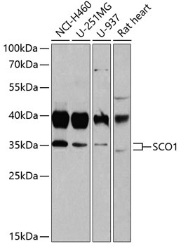
NCI-H460, U-251MG, U-937, Rat heart
Cellular Localization:
Mitochondrion.
Calculated MW:
34kDa
Observed MW:
34kDa
Mammalian cytochrome c oxidase (COX) catalyzes the transfer of reducing equivalents from cytochrome c to molecular oxygen and pumps protons across the inner mitochondrial membrane. In yeast, 2 related COX assembly genes, SCO1 and SCO2 (synthesis of cytochrome c oxidase), enable subunits 1 and 2 to be incorporated into the holoprotein. This gene is the human homolog to the yeast SCO1 gene.
Purification Method
Affinity purification
Gene ID
6341
RRID
AB_2767318
Buffer Information
Store at -20℃. Avoid freeze / thaw cycles. Buffer: PBS containing 50% glycerol, preserved with proclin300 or sodium azide, pH 7.3.
Western blot analysis of various lysates using SCO1 Rabbit pAb (CAB6734) at 1:1000 dilution. Secondary antibody: HRP-conjugated Goat anti-Rabbit IgG (H+L) (CABS014) at 1:10000 dilution. Lysates/proteins: 25μg per lane. Blocking buffer: 3% nonfat dry milk in TBST. Detection: ECL Basic Kit (AbGn00020). Exposure time: 10s.
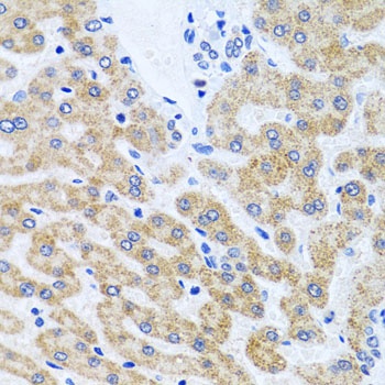
Immunohistochemistry analysis of paraffin-embedded Human liver using SCO1 Rabbit pAb (CAB6734) at dilution of 1:100 (40x lens). Microwave antigen retrieval performed with 0.01M PBS Buffer (pH 7.2) prior to IHC staining.
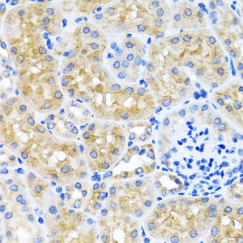
Immunohistochemistry analysis of paraffin-embedded Mouse kidney using SCO1 Rabbit pAb (CAB6734) at dilution of 1:100 (40x lens). Microwave antigen retrieval performed with 0.01M PBS Buffer (pH 7.2) prior to IHC staining.