The SLC3A1 Antibody (CAB5500) is a high-quality antibody developed for reliable detection and analysis of target proteins. This antibody, produced in rabbits, exhibits high specificity and sensitivity for detecting SLC3A1 in human samples, making it ideal for use in Western blot applications.SLC3A1, also known as the heavy subunit of the heterodimeric amino acid transporter system b0,+, plays a crucial role in the uptake of essential amino acids into cells. Dysregulation of SLC3A1 has been implicated in various metabolic disorders and diseases, highlighting the importance of studying its function and expression levels.
This antibody is validated for use in WB, IHC-P, ELISA applications and has demonstrated reactivity against Human, Mouse, Rat samples.
Product Name:
SLC3A1 Antibody
SKU:
CAB5500
Size:
20μL, 100μL
Reactivity:
Human, Mouse, Rat
Conjugate:
Unconjugated
Immunogen:
Recombinant protein (or fragment).This information is considered to be commercially sensitive.
Recommended starting concentration is 1 μg/mL. Please optimize the concentration based on your specific assay requirements.
Synonyms:
D2H, ATR1, NBAT, RBAT, CSNU1, SLC3A1
Positive Sample:
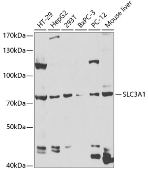
HT-29, HepG2, 293T, BxPC-3, PC-12, Mouse liver
Cellular Localization:
Membrane, Single-Pass Type Ii Membrane Protein.
Calculated MW:
79kDa
Observed MW:
79kDa
This gene encodes a type II membrane glycoprotein which is one of the components of the renal amino acid transporter which transports neutral and basic amino acids in the renal tubule and intestinal tract. Mutations and deletions in this gene are associated with cystinuria. Alternatively spliced transcript variants have been described, but their biological validity has not been determined.
Purification Method
Affinity purification
Gene ID
6519
RRID
AB_2766296
Buffer Information
Store at -20℃. Avoid freeze / thaw cycles. Buffer: PBS containing 50% glycerol, preserved with proclin300 or sodium azide, pH 7.3.
Western blot analysis of various lysates using SLC3A1 Rabbit pAb (CAB5500) at 1:1000 dilution. Secondary antibody: HRP-conjugated Goat anti-Rabbit IgG (H+L) (CABS014) at 1:10000 dilution. Lysates/proteins: 25μg per lane. Blocking buffer: 3% nonfat dry milk in TBST. Detection: ECL Basic Kit (AbGn00020). Exposure time: 90s.
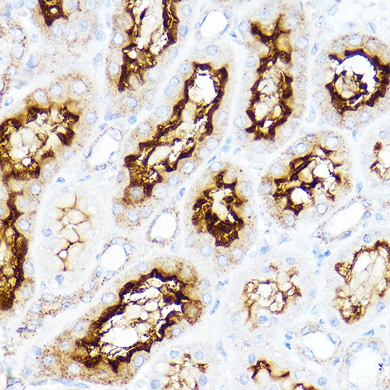
Immunohistochemistry analysis of paraffin-embedded Rat kidney using SLC3A1 Rabbit pAb (CAB5500) at dilution of 1:200 (40x lens). High pressure antigen retrieval performed with 0.01M Citrate buffer (pH 6.0) prior to IHC staining.