The ST8SIA1 Antibody (CAB9648) is a high-quality antibody developed for reliable detection and analysis of target proteins. This antibody, generated in rabbits, is highly specific for human samples and has been validated for use in Western blotting applications. By targeting the ST8SIA1 enzyme, this antibody allows for the detection and analysis of ST8SIA1 expression in a variety of cell types, making it ideal for research in neurobiology, developmental biology, and cancer biology.
This antibody is validated for use in WB, ELISA applications and has demonstrated reactivity against Mouse samples.
Product Name:
ST8SIA1 Antibody
SKU:
CAB9648
Size:
20μL, 100μL
Reactivity:
Mouse
Conjugate:
Unconjugated
Immunogen:
Recombinant protein (or fragment).This information is considered to be commercially sensitive.
Recommended starting concentration is 1 μg/mL. Please optimize the concentration based on your specific assay requirements.
Synonyms:
GD3S, SIAT8, SIAT8A, SIAT8-A, ST8SiaI, ST8SIA1
Positive Sample:
Mouse kidney
Cellular Localization:
Golgi Apparatus Membrane, Single-Pass Type Ii Membrane Protein.
Calculated MW:
41kDa
Observed MW:
41kDa
Gangliosides are membrane-bound glycosphingolipids containing sialic acid. Ganglioside GD3 is known to be important for cell adhesion and growth of cultured malignant cells. The protein encoded by this gene is a type II membrane protein that catalyzes the transfer of sialic acid from CMP-sialic acid to GM3 to produce gangliosides GD3 and GT3. The encoded protein may be found in the Golgi apparatus and is a member of glycosyltransferase family 29. Alternatively spliced transcript variants have been found for this gene.
Purification Method
Affinity purification
Gene ID
6489
RRID
AB_2772414
Buffer Information
Store at -20℃. Avoid freeze / thaw cycles. Buffer: PBS containing 50% glycerol, preserved with proclin300 or sodium azide, pH 7.3.
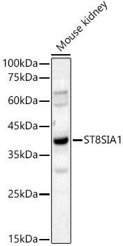
Western blot analysis of lysates from Mouse kidney, using ST8SIA1 Rabbit pAb (CAB9648) at 1:8000 dilution. Secondary antibody: HRP-conjugated Goat anti-Rabbit IgG (H+L) (CABS014) at 1:10000 dilution. Lysates/proteins: 25μg per lane. Blocking buffer: 3% nonfat dry milk in TBST. Detection: ECL Basic Kit (AbGn00020). Exposure time: 90s.