The TBC1D4 Antibody (CAB18216) is a high-quality antibody developed for reliable detection and analysis of target proteins. This antibody, generated in rabbits, is highly specific to human samples and has been extensively validated for use in Western blotting applications. By binding to the TBC1D4 protein, this antibody allows for the detection and analysis of TBC1D4 expression in a variety of cell types, making it ideal for studies in diabetes, metabolism, and endocrinology.TBC1D4, also known as AS160, plays a crucial role in the translocation of glucose transporter proteins to the cell membrane in response to insulin signaling, thereby regulating glucose uptake and metabolism.
This antibody is validated for use in WB, IF/ICC, ELISA applications and has demonstrated reactivity against Mouse, Rat samples.
Product Name:
TBC1D4 Antibody
SKU:
CAB18216
Size:
20μL, 100μL
Reactivity:
Mouse, Rat
Immunogen:
Recombinant protein (or fragment).This information is considered to be commercially sensitive.
Recommended starting concentration is 1 μg/mL. Please optimize the concentration based on your specific assay requirements.
Synonyms:
AS160, NIDDM5, TBC1D4
Positive Sample:
Rat skeletal muscle
Cellular Localization:
Cytosol.
Calculated MW:
147kDa
Observed MW:
160kDa
This gene is a member of the Tre-2/BUB2/CDC16 domain family. The protein encoded by this gene is a Rab-GTPase-activating protein, and contains two phopshotyrosine-binding domains (PTB1 and PTB2), a calmodulin-binding domain (CBD), a Rab-GTPase domain, and multiple AKT phosphomotifs. This protein is thought to play an important role in glucose homeostasis by regulating the insulin-dependent trafficking of the glucose transporter 4 (GLUT4), important for removing glucose from the bloodstream into skeletal muscle and fat tissues. Reduced expression of this gene results in an increase in GLUT4 levels at the plasma membrane, suggesting that this protein is important in intracellular retention of GLUT4 under basal conditions. When exposed to insulin, this protein is phosphorylated, dissociates from GLUT4 vesicles, resulting in increased GLUT4 at the cell surface, and enhanced glucose transport. Phosphorylation of this protein by AKT is required for proper translocation of GLUT4 to the cell surface. Individuals homozygous for a mutation in this gene are at higher risk for type 2 diabetes and have higher levels of circulating glucose and insulin levels after glucose ingestion. Alternative splicing results in multiple transcript variants encoding different isoforms.
Purification Method
Affinity purification
Gene ID
9882
RRID
AB_2861992
Buffer Information
Store at -20℃. Avoid freeze / thaw cycles. Buffer: PBS with 0.01% thimerosal,50% glycerol,pH7.3.
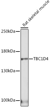
Western blot analysis of lysates from Rat skeletal muscle, using TBC1D4 Rabbit pAb (CAB18216) at 1:1000 dilution. Secondary antibody: HRP-conjugated Goat anti-Rabbit IgG (H+L) (CABS014) at 1:10000 dilution. Lysates/proteins: 25μg per lane. Blocking buffer: 3% nonfat dry milk in TBST. Detection: ECL Basic Kit (AbGn00020). Exposure time: 30s.
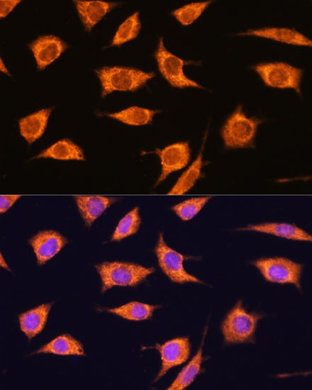
Immunofluorescence analysis of L929 cells using TBC1D4 Rabbit pAb (CAB18216) at dilution of 1:100. Secondary antibody: Cy3-conjugated Goat anti-Rabbit IgG (H+L) (CABS007) at 1:500 dilution. Blue: DAPI for nuclear staining.