The Thioredoxin 1 (Trx1/TXN) Antibody (CAB7638) is a high-quality antibody developed for reliable detection and analysis of target proteins. This antibody, raised in rabbits, exhibits high reactivity with human samples and has been validated for use in Western blot applications. By binding specifically to Thioredoxin, this antibody enables accurate detection and analysis of the protein in various cell types.Thioredoxin is essential for maintaining cellular redox balance and has been implicated in a wide range of physiological processes, including cell growth, apoptosis, and immune responses.
This antibody is validated for use in WB, IHC-P, IF/ICC, IP, ELISA applications and has demonstrated reactivity against Human, Mouse samples.
Product Name:
Thioredoxin 1 (Trx1/TXN) Antibody
SKU:
CAB7638
Size:
20μL, 100μL
Reactivity:
Human, Mouse
Conjugate:
Unconjugated
Immunogen:
Synthetic peptide. This information is considered to be commercially sensitive.
0.5μg-4μg antibody for 200μg-400μg extracts of whole cells
ELISA
Recommended starting concentration is 1 μg/mL. Please optimize the concentration based on your specific assay requirements.
Synonyms:
TRX, TRDX, TRX1, Trx80, Thioredoxin 1 (Trx1/TXN)
Positive Sample:
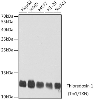
HepG2, H460, MCF7, HT-29, SKOV3
Cellular Localization:
Cytoplasm, Nucleus, Secreted.
Calculated MW:
12kDa
Observed MW:
12kDa
The protein encoded by this gene acts as a homodimer and is involved in many redox reactions. The encoded protein is active in the reversible S-nitrosylation of cysteines in certain proteins, which is part of the response to intracellular nitric oxide. This protein is found in the cytoplasm. Two transcript variants encoding different isoforms have been found for this gene.
Purification Method
Affinity purification
Gene ID
7295
RRID
AB_2768150
Buffer Information
Store at -20℃. Avoid freeze / thaw cycles. Buffer: PBS containing 50% glycerol, preserved with proclin300 or sodium azide, pH 7.3.
Western blot analysis of various lysates using Thioredoxin 1 (Trx1/TXN) Rabbit pAb (CAB7638) at 1:1000 dilution. Secondary antibody: HRP-conjugated Goat anti-Rabbit IgG (H+L) (CABS014) at 1:10000 dilution. Lysates/proteins: 25μg per lane. Blocking buffer: 3% nonfat dry milk in TBST. Detection: ECL Basic Kit (AbGn00020). Exposure time: 90s.
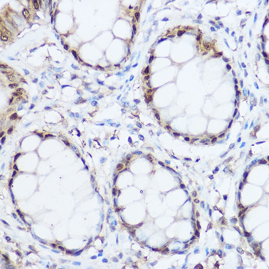
Immunohistochemistry analysis of paraffin-embedded Human colon using Thioredoxin 1 (Trx1/Thioredoxin 1 (Trx1/TXN)) Rabbit pAb (CAB7638) at dilution of 1:300 (40x lens). High pressure antigen retrieval performed with 0.01M Citrate buffer (pH 6.0) prior to IHC staining.
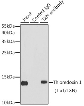
Immunoprecipitation analysis of 150 μg extracts of MCF7 cells using 3 μg Thioredoxin 1 (Trx1/Thioredoxin 1 (Trx1/TXN)) antibody (CAB7638). Western blot was performed from the immunoprecipitate using Thioredoxin 1 (Trx1/Thioredoxin 1 (Trx1/TXN)) antibody (CAB7638) at a dilution of 1:500.


 ELISA Kit (AEFI00818)")
 ELISA Kit (AEFI00818)")


 ELISA Kit (PREB0175)")
")