The TIA1 Antibody (CAB12523) is a high-quality antibody developed for reliable detection and analysis of target proteins. Raised in rabbits, this antibody is highly reactive with human samples and is validated for use in Western blot applications. By binding to the TIA1 protein, this antibody allows for the detection and analysis of TIA1 in various cell types, making it ideal for studies in molecular biology, RNA metabolism, and neurodegenerative diseases.
This antibody is validated for use in WB, IF/ICC, ELISA applications and has demonstrated reactivity against Human, Mouse samples.
Product Name:
TIA1 Antibody
SKU:
CAB12523
Size:
20μL, 100μL
Reactivity:
Human, Mouse
Conjugate:
Unconjugated
Immunogen:
Synthetic peptide. This information is considered to be commercially sensitive.
Recommended starting concentration is 1 μg/mL. Please optimize the concentration based on your specific assay requirements.
Synonyms:
WDM, ALS26, TIA-1, TIA1
Positive Sample:
Jurkat
Cellular Localization:
Cytoplasmic Granule, Nucleus.
Calculated MW:
43kDa
Observed MW:
44kDa
The product encoded by this gene is a member of a RNA-binding protein family and possesses nucleolytic activity against cytotoxic lymphocyte (CTL) target cells. It has been suggested that this protein may be involved in the induction of apoptosis as it preferentially recognizes poly(A) homopolymers and induces DNA fragmentation in CTL targets. The major granule-associated species is a 15-kDa protein that is thought to be derived from the carboxyl terminus of the 40-kDa product by proteolytic processing. Alternative splicing resulting in different isoforms has been found for this gene.
Purification Method
Affinity purification
Gene ID
7072
RRID
AB_2759363
Buffer Information
Store at -20℃. Avoid freeze / thaw cycles. Buffer: PBS containing 50% glycerol, preserved with proclin300 or sodium azide, pH 7.3.
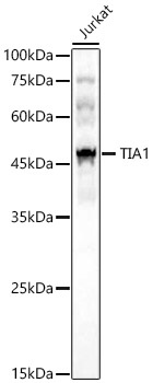
Western blot analysis of lysates from Jurkat cells, using TIA1 Rabbit pAb (CAB12523) at 1:1000 dilution. Secondary antibody: HRP-conjugated Goat anti-Rabbit IgG (H+L) (CABS014) at 1:10000 dilution. Lysates/proteins: 25μg per lane. Blocking buffer: 3% nonfat dry milk in TBST. Detection: ECL Basic Kit (AbGn00020). Exposure time: 60s.
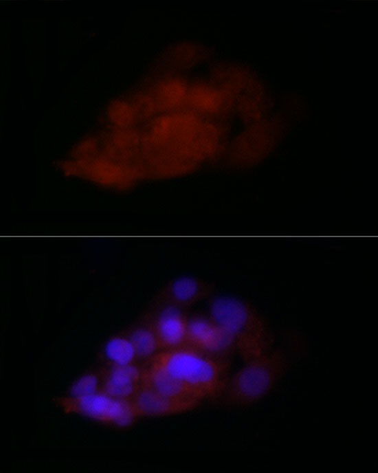
Immunofluorescence analysis of HepG2 cells using TIA1 Rabbit pAb (CAB12523) at dilution of 1:100 (40x lens). Secondary antibody: Cy3-conjugated Goat anti-Rabbit IgG (H+L) (CABS007) at 1:500 dilution. Blue: DAPI for nuclear staining.