The TLR2 Monoclonal Antibody (CAB19125) is a high-quality antibody developed for reliable detection and analysis of target proteins. This high-quality antibody, produced in rabbits, is highly specific for TLR2 and has been validated for use in various applications, including Western blotting and immunofluorescence.TLR2 is a key player in the innate immune response, recognizing components of bacteria, viruses, and fungi, and activating downstream signaling pathways that lead to inflammation and host defense.
This antibody is validated for use in WB, IF/ICC, ELISA, IF-P applications and has demonstrated reactivity against Human, Mouse samples.
Product Name:
TLR2 Monoclonal Antibody
SKU:
CAB19125
Size:
20μL, 100μL
Reactivity:
Human, Mouse
Clone Number:
ARC0433
Conjugate:
Unconjugated
Immunogen:
Recombinant protein (or fragment).This information is considered to be commercially sensitive.
Recommended starting concentration is 1 μg/mL. Please optimize the concentration based on your specific assay requirements.
Synonyms:
TIL4, CD282, TLR2
Positive Sample:
THP-1
Cellular Localization:
Membrane, Single-Pass Type I Membrane Protein.
Calculated MW:
90kDa
Observed MW:
90-105kDa
The protein encoded by this gene is a member of the Toll-like receptor (TLR) family which plays a fundamental role in pathogen recognition and activation of innate immunity. TLRs are highly conserved from Drosophila to humans and share structural and functional similarities. This protein is a cell-surface protein that can form heterodimers with other TLR family members to recognize conserved molecules derived from microorganisms known as pathogen-associated molecular patterns (PAMPs). Activation of TLRs by PAMPs leads to an up-regulation of signaling pathways to modulate the host's inflammatory response. This gene is also thought to promote apoptosis in response to bacterial lipoproteins. This gene has been implicated in the pathogenesis of several autoimmune diseases. Alternative splicing results in multiple transcript variants.
Purification Method
Affinity purification
Gene ID
7097
RRID
AB_2862618
Buffer Information
Store at -20℃. Avoid freeze / thaw cycles. Buffer: PBS containing 50% glycerol and 0.05% BSA, preserved with proclin300 or sodium azide, pH 7.3.
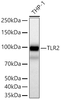
Western blot analysis of lysates from THP-1 cells using TLR2 Rabbit mAb (CAB19125) at 1:1000 dilution incubated overnight at 4℃. Secondary antibody: HRP-conjugated Goat anti-Rabbit IgG (H+L) (CABS014) at 1:10000 dilution. Lysates/proteins: 25 μg per lane. Blocking buffer: 3% nonfat dry milk in TBST. Detection: ECL Basic Kit (AbGn00020). Exposure time: 45s.
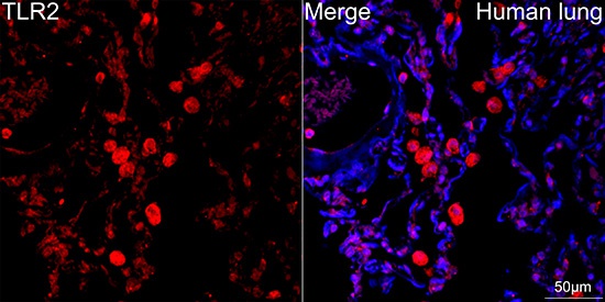
Confocal imaging of paraffin-embedded Human lung tissue using TLR2 Rabbit mAb (CAB19125, dilution 1:100) followed by a further incubation with Cy3 Goat Anti-Rabbit IgG (H+L) (CABS007, dilution 1:500) (Red). DAPI was used for nuclear staining (Blue). High pressure antigen retrieval performed with 0.01M Citrate Buffer (pH 6.0) prior to IF staining. Objective: 40x.
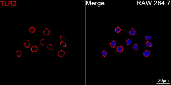
Confocal imaging of RAW 264.7 cells using TLR2 Rabbit mAb (CAB19125, dilution 1:200) followed by a further incubation with Cy3 Goat Anti-Rabbit IgG (H+L) (CABS007, dilution 1:500) (Red). DAPI was used for nuclear staining (Blue). Objective: 100x.


![Anti-TLR2 [R06-8B6] Monoclonal Antibody (AGMB00156)](https://cdn11.bigcommerce.com/s-h68l9z2lnx/images/stencil/590x590/products/271445/694538/anti-tlr2-r06-8b6-monoclonal-antibody-agmb00156__81949.1774512801.jpg?c=2 "Anti-TLR2 [R06-8B6] Monoclonal Antibody (AGMB00156)")
![Anti-TLR2 [R08-7F2] Monoclonal Antibody (AGMB04108)](https://cdn11.bigcommerce.com/s-h68l9z2lnx/images/stencil/590x590/products/275397/678603/anti-tlr2-r08-7f2-monoclonal-antibody-agmb04108__61267.1773036270.jpg?c=2 "Anti-TLR2 [R08-7F2] Monoclonal Antibody (AGMB04108)")


