The TNFRSF10A Antibody (CAB12540) is a high-quality antibody developed for reliable detection and analysis of target proteins. This antibody, generated in rabbits, exhibits high reactivity with human samples and is validated for use in Western blot applications, enabling the detection and analysis of TNFRSF10A protein in various cell types.TNFRSF10A, also known as DR4 or TRAIL-R1, is a receptor involved in the apoptotic pathway, making it a key player in controlling cell survival and death. Its dysregulation has been implicated in various diseases, including cancer, making it a promising target for therapeutic intervention.
This antibody is validated for use in WB, IF/ICC, ELISA, IF-P applications and has demonstrated reactivity against Human, Mouse, Rat samples.
Product Name:
TNFRSF10A Antibody
SKU:
CAB12540
Size:
20μL, 100μL
Reactivity:
Human, Mouse, Rat
Conjugate:
Unconjugated
Immunogen:
Recombinant protein (or fragment).This information is considered to be commercially sensitive.
Recommended starting concentration is 1 μg/mL. Please optimize the concentration based on your specific assay requirements.
Synonyms:
DR4, APO2, CD261, TRAILR1, TRAILR-1, TNFRSF10A
Positive Sample:
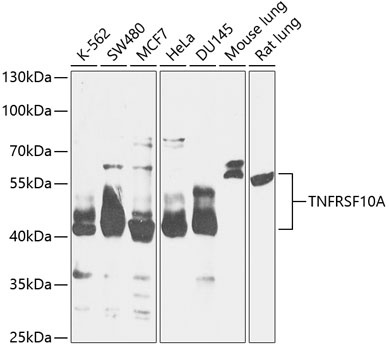
K-562, SW480, MCF7, HeLa, DU145, Mouse lung, Rat lung
Cellular Localization:
Membrane, Single-Pass Type I Membrane Protein.
Calculated MW:
50kDa
Observed MW:
40-55kDa
The protein encoded by this gene is a member of the TNF-receptor superfamily. This receptor is activated by tumor necrosis factor-related apoptosis inducing ligand (TNFSF10/TRAIL), and thus transduces cell death signal and induces cell apoptosis. Studies with FADD-deficient mice suggested that FADD, a death domain containing adaptor protein, is required for the apoptosis mediated by this protein.
Purification Method
Affinity purification
Gene ID
8797
RRID
AB_2759380
Buffer Information
Store at -20℃. Avoid freeze / thaw cycles. Buffer: PBS containing 50% glycerol, preserved with proclin300 or sodium azide, pH 7.3.
Western blot analysis of various lysates using TNFRSF10A Rabbit pAb (CAB12540) at 1:1000 dilution. Secondary antibody: HRP-conjugated Goat anti-Rabbit IgG (H+L) (CABS014) at 1:10000 dilution. Lysates/proteins: 25μg per lane. Blocking buffer: 3% nonfat dry milk in TBST. Detection: ECL Basic Kit (AbGn00020). Exposure time: 60s.
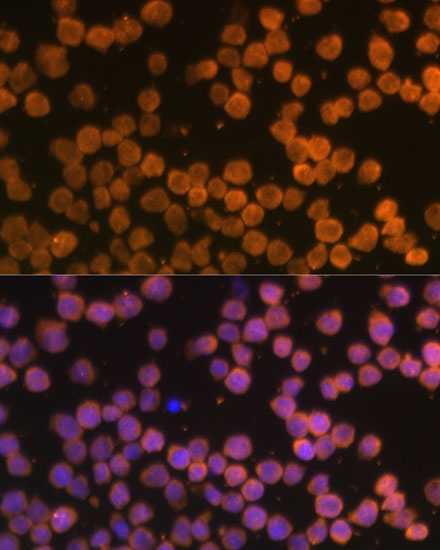
Immunofluorescence analysis of THP-1 cells using TNFRSF10A Rabbit pAb (CAB12540) at dilution of 1:100 (40x lens). Secondary antibody: Cy3-conjugated Goat anti-Rabbit IgG (H+L) (CABS007) at 1:500 dilution. Blue: DAPI for nuclear staining.