The OX40L Antibody (CAB18389) is a high-quality antibody developed for reliable detection and analysis of target proteins. This rabbit-derived antibody is highly specific to human TNFSF4 and is validated for use in Western blot applications. It binds specifically to TNFSF4, allowing for accurate detection and analysis in various cell types.TNFSF4, also known as OX40 ligand, plays a crucial role in regulating immune responses by activating T cells and promoting cytokine production. Its involvement in inflammatory processes makes it a key target for research in immunology and autoimmune diseases.
This antibody is validated for use in WB, IF/ICC, ELISA applications and has demonstrated reactivity against Human, Mouse samples.
Product Name:
OX40L Antibody
SKU:
CAB18389
Size:
20μL, 100μL
Reactivity:
Human, Mouse
Immunogen:
Recombinant protein (or fragment).This information is considered to be commercially sensitive.
This gene encodes a cytokine of the tumor necrosis factor (TNF) ligand family. The encoded protein functions in T cell antigen-presenting cell (APC) interactions and mediates adhesion of activated T cells to endothelial cells. Polymorphisms in this gene have been associated with Sjogren's syndrome and systemic lupus erythematosus. Alternative splicing results in multiple transcript variants.
Purification Method
Affinity purification
Gene ID
7292
RRID
AB_2862156
Buffer Information
Store at -20℃. Avoid freeze / thaw cycles. Buffer: PBS containing 50% glycerol, preserved with proclin300 or sodium azide, pH 7.3.
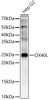
Western blot analysis of lysates from Hep G2 cells using OX40L Rabbit pAb (CAB18389) at 1:500 dilution. Secondary antibody: HRP-conjugated Goat anti-Rabbit IgG (H+L) (CABS014) at 1:10000 dilution. Lysates/proteins: 25 μg per lane. Blocking buffer: 3% nonfat dry milk in TBST. Detection: ECL Enhanced Kit (AbGn00021). Exposure time: 60s.
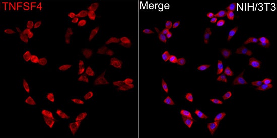
Immunofluorescence analysis of NIH/3T3 cells using OX40L Rabbit pAb (CAB18389) at dilution of 1:50 (40x lens). Secondary antibody: Cy3-conjugated Goat anti-Rabbit IgG (H+L) (CABS007) at 1:500 dilution. Blue: DAPI for nuclear staining.