DNA topoisomerase I (TOP1) Monoclonal Antibody (CAB12409)
The DNA topoisomerase I (TOP1) Monoclonal Antibody (CAB12409) is a high-quality antibody developed for reliable detection and analysis of target proteins. This highly specific antibody, produced in rabbits, exhibits strong reactivity with human samples and is validated for use in Western blot and immunohistochemistry applications.TOP1 plays a crucial role in maintaining genomic stability and is a known target for cancer therapies, making it a key focus in cancer research. By targeting TOP1, researchers can gain valuable insights into the mechanisms underlying cancer development and potentially identify novel therapeutic strategies.
This antibody is validated for use in WB, IHC-P, IF/ICC, IP, ELISA applications and has demonstrated reactivity against Human, Mouse, Rat samples.
Product Name:
DNA topoisomerase I (TOP1) Monoclonal Antibody
SKU:
CAB12409
Size:
20μL, 100μL
Reactivity:
Human, Mouse, Rat
Clone Number:
ARC0708
Conjugate:
Unconjugated
Immunogen:
Synthetic peptide. This information is considered to be commercially sensitive.
0.5μg-4μg antibody for 200μg-400μg extracts of whole cells
ELISA
Recommended starting concentration is 1 μg/mL. Please optimize the concentration based on your specific assay requirements.
Synonyms:
TOPI, DNA topoisomerase I (TOP1)
Positive Sample:
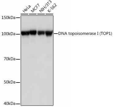
Mouse thymus, HeLa, MCF7, NIH/3T3, K-562
Cellular Localization:
Nucleus, Nucleolus, Nucleoplasm.
Calculated MW:
91kDa
Observed MW:
100kDa
This gene encodes a DNA topoisomerase, an enzyme that controls and alters the topologic states of DNA during transcription. This enzyme catalyzes the transient breaking and rejoining of a single strand of DNA which allows the strands to pass through one another, thus altering the topology of DNA. This gene is localized to chromosome 20 and has pseudogenes which reside on chromosomes 1 and 22.
Purification Method
Affinity purification
Gene ID
7150
RRID
AB_2861661
Buffer Information
Store at -20℃. Avoid freeze / thaw cycles. Buffer: PBS containing 50% glycerol and 0.05% BSA, preserved with proclin300 or sodium azide, pH 7.3.
Western blot analysis of various lysates using DNA topoisomerase I (DNA topoisomerase I (TOP1)) Rabbit mAb (CAB12409) at 1:1000 dilution. Secondary antibody: HRP-conjugated Goat anti-Rabbit IgG (H+L) (CABS014) at 1:10000 dilution. Lysates/proteins: 25μg per lane. Blocking buffer: 3% nonfat dry milk in TBST. Detection: ECL Basic Kit (AbGn00020). Exposure time: 1s.
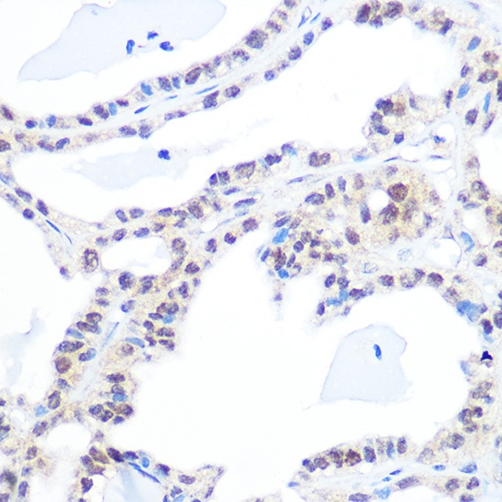
Immunohistochemistry analysis of paraffin-embedded Human thyroid cancer using DNA topoisomerase I (DNA topoisomerase I (TOP1)) Rabbit mAb (CAB12409) at dilution of 1:100 (40x lens). Microwave antigen retrieval performed with 0.01M PBS Buffer (pH 7.2) prior to IHC staining.
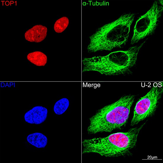
Confocal imaging of U-2 OS cells using DNA topoisomerase I (TOP1) Rabbit mAb (CAB12409,dilution 1:100)(Red). The cells were counterstained with α-Tubulin Mouse mAb (AC012,dilution 1:400) (Green). DAPI was used for nuclear staining (blue). Objective: 100x.
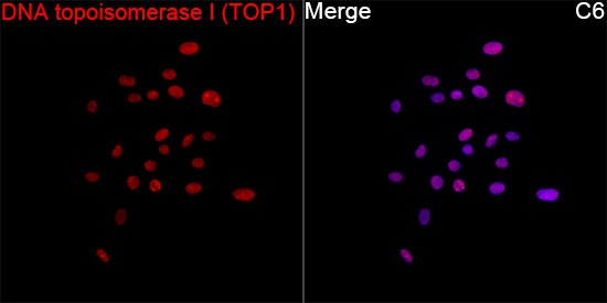
Immunofluorescence analysis of C6 cells using DNA topoisomerase I (TOP1) Rabbit mAb (CAB12409) at a dilution of 1:100 (40x lens). Secondary antibody: Cy3-conjugated Goat anti-Rabbit IgG (H+L)(CABS007) at 1:500 dilution. Blue: DAPI for nuclear staining.
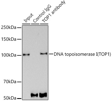
Immunoprecipitation analysis of 300 μg extracts of MCF7 cells using 3 μg DNA topoisomerase I (TOP1) antibody (CAB12409). Western blot was performed from the immunoprecipitate using DNA topoisomerase I (TOP1) antibody (CAB12409) at a dilution of 1:1000.


 ELISA Kit (HUEB2675)")
 ELISA Kit (RTEB1676)")
 ELISA Kit (MOEB2421)")