The TRPV6 Antibody (CAB16128) is a high-quality antibody developed for reliable detection and analysis of target proteins. This antibody, produced in rabbits, is highly specific to human TRPV6 and has been validated for use in Western blot applications. By specifically binding to TRPV6, researchers can detect and analyze the protein in various cell types, making it an excellent tool for investigations in calcium signaling, cancer biology, and reproductive health.
This antibody is validated for use in WB, IHC-P, ELISA applications and has demonstrated reactivity against Human, Mouse, Rat samples.
Product Name:
TRPV6 Antibody
SKU:
CAB16128
Size:
20μL, 100μL
Reactivity:
Human, Mouse, Rat
Conjugate:
Unconjugated
Immunogen:
Synthetic peptide. This information is considered to be commercially sensitive.
This gene encodes a member of a family of multipass membrane proteins that functions as calcium channels. The encoded protein contains N-terminal ankyrin repeats, which are required for channel assembly and regulation. Translation initiation for this protein occurs at a non-AUG start codon that is decoded as methionine. This gene is situated next to a closely related gene for transient receptor potential cation channel subfamily V member 5 (TRPV5). This locus has experienced positive selection in non-African populations, resulting in several non-synonymous codon differences among individuals of different genetic backgrounds.
Purification Method
Affinity purification
Gene ID
55503
RRID
AB_2763573
Buffer Information
Store at -20℃. Avoid freeze / thaw cycles. Buffer: PBS containing 50% glycerol, preserved with proclin300 or sodium azide, pH 7.3.
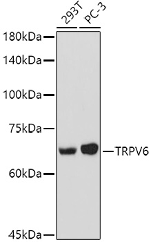
Western blot analysis of various lysates using TRPV6 Rabbit pAb (CAB16128) at 1:1000 dilution. Secondary antibody: HRP-conjugated Goat anti-Rabbit IgG (H+L) (CABS014) at 1:10000 dilution. Lysates/proteins: 25μg per lane. Blocking buffer: 3% nonfat dry milk in TBST. Detection: ECL Basic Kit (AbGn00020). Exposure time: 90s.
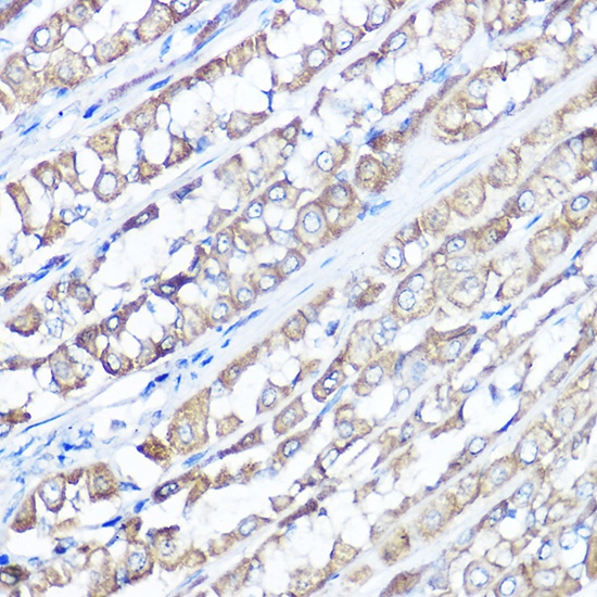
Immunohistochemistry analysis of paraffin-embedded Rat stomach using TRPV6 Rabbit pAb (CAB16128) at dilution of 1:100 (40x lens). High pressure antigen retrieval performed with 0.01M Citrate buffer (pH 6.0) prior to IHC staining.