The VCP Monoclonal Antibody (CAB1402) is a high-quality antibody developed for reliable detection and analysis of target proteins. Raised in rabbits, this antibody offers high specificity and sensitivity for detecting VCP in human samples, making it an essential tool for researchers in cell biology and cancer research.VCP, also known as p97, is a AAA+ ATPase enzyme involved in multiple cellular pathways, including endoplasmic reticulum-associated degradation (ERAD) and autophagy. Dysregulation of VCP has been linked to various diseases, including neurodegenerative disorders and cancer.
This antibody is validated for use in WB, IF/ICC, ELISA applications and has demonstrated reactivity against Human, Mouse, Rat samples.
Product Name:
VCP Monoclonal Antibody
SKU:
CAB1402
Size:
20μL, 100μL
Reactivity:
Human, Mouse, Rat
Clone Number:
ARC0728
Conjugate:
Unconjugated
Immunogen:
Synthetic peptide. This information is considered to be commercially sensitive.
This gene encodes a member of the AAA ATPase family of proteins. The encoded protein plays a role in protein degradation, intracellular membrane fusion, DNA repair and replication, regulation of the cell cycle, and activation of the NF-kappa B pathway. This protein forms a homohexameric complex that interacts with a variety of cofactors and extracts ubiquitinated proteins from lipid membranes or protein complexes. Mutations in this gene cause IBMPFD (inclusion body myopathy with paget disease of bone and frontotemporal dementia), ALS (amyotrophic lateral sclerosis) and Charcot-Marie-Tooth disease in human patients.
Purification Method
Affinity purification
Gene ID
7415
RRID
AB_2861698
Buffer Information
Store at -20℃. Avoid freeze / thaw cycles. Buffer: PBS containing 50% glycerol and 0.05% BSA, preserved with proclin300 or sodium azide, pH 7.3.
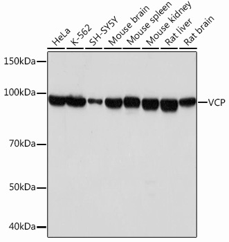
Western blot analysis of various lysates using VCP Rabbit mAb (CAB1402) at 1:1000 dilution. Secondary antibody: HRP-conjugated Goat anti-Rabbit IgG (H+L) (CABS014) at 1:10000 dilution. Lysates/proteins: 25μg per lane. Blocking buffer: 3% nonfat dry milk in TBST. Detection: ECL Basic Kit (AbGn00020). Exposure time: 10s.
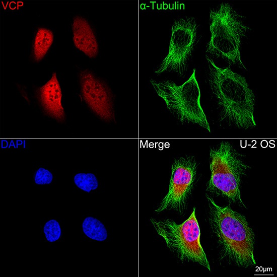
Confocal imaging of U-2 OS cells using VCP Rabbit mAb (CAB1402, dilution 1:200) followed by a further incubation with Cy3 Goat Anti-Rabbit IgG (H+L) (CABS007, dilution 1:500) (Red). The cells were counterstained with α-Tubulin Mouse mAb (AC012, dilution 1:400) followed by incubation with ABflo® 488-conjugated Goat Anti-Mouse IgG (H+L) Ab (CABS076, dilution 1:500) (Green). DAPI was used for nuclear staining (Blue). Objective: 100x.
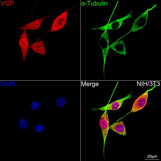
Confocal imaging of NIH/3T3 cells using VCP Rabbit mAb (CAB1402, dilution 1:200) followed by a further incubation with Cy3 Goat Anti-Rabbit IgG (H+L) (CABS007, dilution 1:500) (Red). The cells were counterstained with α-Tubulin Mouse mAb (AC012, dilution 1:400) followed by incubation with ABflo® 488-conjugated Goat Anti-Mouse IgG (H+L) Ab (CABS076, dilution 1:500) (Green). DAPI was used for nuclear staining (Blue). Objective: 100x.


![Anti-VCP [R06-9D7] Monoclonal Antibody (AGMB01215)](https://cdn11.bigcommerce.com/s-h68l9z2lnx/images/stencil/590x590/products/272504/693027/anti-vcp-r06-9d7-monoclonal-antibody-agmb01215__84207.1774508007.jpg?c=2 "Anti-VCP [R06-9D7] Monoclonal Antibody (AGMB01215)")


