The ACVRL1 Monoclonal Antibody (CAB9755) is a high-quality antibody developed for reliable detection and analysis of target proteins. This antibody, raised in rabbits, is highly specific to human samples and has been validated for use in Western blot applications.ACVRL1 is a type I receptor for the TGF-beta superfamily of ligands and plays a critical role in regulating blood vessel formation and maintaining vascular integrity. Dysregulation of ACVRL1 has been linked to diseases such as hereditary hemorrhagic telangiectasia (HHT) and various cancers.
This antibody is validated for use in WB, IF/ICC, ELISA applications and has demonstrated reactivity against Human, Mouse, Rat samples.
Product Name:
ACVRL1 Monoclonal Antibody
SKU:
CAB9755
Size:
20μL, 100μL
Reactivity:
Human, Mouse, Rat
Clone Number:
ARC1735
Conjugate:
Unconjugated
Immunogen:
Synthetic peptide. This information is considered to be commercially sensitive.
This gene encodes a type I cell-surface receptor for the TGF-beta superfamily of ligands. It shares with other type I receptors a high degree of similarity in serine-threonine kinase subdomains, a glycine- and serine-rich region (called the GS domain) preceding the kinase domain, and a short C-terminal tail. The encoded protein, sometimes termed ALK1, shares similar domain structures with other closely related ALK or activin receptor-like kinase proteins that form a subfamily of receptor serine/threonine kinases. Mutations in this gene are associated with hemorrhagic telangiectasia type 2, also known as Rendu-Osler-Weber syndrome 2.
Purification Method
Affinity purification
Gene ID
94
RRID
AB_2863773
Buffer Information
Store at -20℃. Avoid freeze / thaw cycles. Buffer: PBS containing 50% glycerol and 0.05% BSA, preserved with proclin300 or sodium azide, pH 7.3.
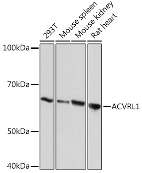
Western blot analysis of various lysates using ACVRL1 Rabbit mAb (CAB9755) at 1:1000 dilution. Secondary antibody: HRP-conjugated Goat anti-Rabbit IgG (H+L) (CABS014) at 1:10000 dilution. Lysates/proteins: 25μg per lane. Blocking buffer: 3% nonfat dry milk in TBST. Detection: ECL Basic Kit (AbGn00020). Exposure time: 10s.
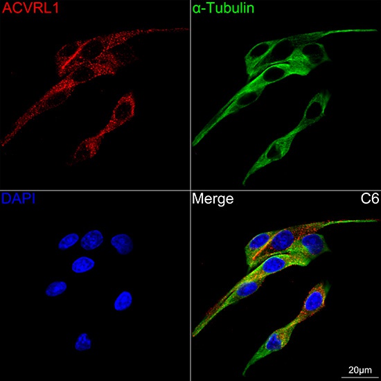
Confocal imaging of C6 cells using ACVRL1 Rabbit mAb (CAB9755, dilution 1:100) followed by a further incubation with Cy3 Goat Anti-Rabbit IgG (H+L) (CABS007, dilution 1:500) (Red). The cells were counterstained with α-Tubulin Mouse mAb (AC012, dilution 1:400) followed by incubation with ABflo® 488-conjugated Goat Anti-Mouse IgG (H+L) Ab (CABS076, dilution 1:500) (Green). DAPI was used for nuclear staining (Blue). Objective: 100x.