The Musashi-1 (MSI1) Monoclonal Antibody (CAB9122) is a high-quality antibody developed for reliable detection and analysis of target proteins. This antibody, produced in rabbits, is highly specific for detecting Musashi-1 in human samples and has been validated for use in Western blot and immunohistochemistry applications. By binding to Musashi-1, this antibody enables researchers to investigate the expression and localization of this important protein in various tissues and cell types.Musashi-1 is known for its role in promoting self-renewal and maintaining the pluripotency of stem cells, making it a crucial target for studies in stem cell biology and regenerative medicine.
This antibody is validated for use in WB, IF/ICC, ELISA applications and has demonstrated reactivity against Human, Mouse samples.
Product Name:
Musashi-1 (MSI1) Monoclonal Antibody
SKU:
CAB9122
Size:
20μL, 100μL
Reactivity:
Human, Mouse
Clone Number:
ARC1796
Conjugate:
Unconjugated
Immunogen:
Synthetic peptide. This information is considered to be commercially sensitive.
Recommended starting concentration is 1 μg/mL. Please optimize the concentration based on your specific assay requirements.
Synonyms:
MSI1, musashi RNA binding protein 1, Musashi-1 (MSI1)
Positive Sample:
SH-SY5Y, Mouse brain
Cellular Localization:
Cytoplasm, Nucleus.
Calculated MW:
39kDa
Observed MW:
39kDa
This gene encodes a protein containing two conserved tandem RNA recognition motifs. Similar proteins in other species function as RNA-binding proteins and play central roles in posttranscriptional gene regulation. Expression of this gene has been correlated with the grade of the malignancy and proliferative activity in gliomas and melanomas. A pseudogene for this gene is located on chromosome 11q13.
Purification Method
Affinity purification
Gene ID
4440
RRID
AB_2863658
Buffer Information
Store at -20℃. Avoid freeze / thaw cycles. Buffer: PBS containing 50% glycerol and 0.05% BSA, preserved with proclin300 or sodium azide, pH 7.3.
Western blot analysis of various lysates using Musashi-1 (MSI1) Rabbit mAb (CAB9122) at 1:1000 dilution. Secondary antibody: HRP-conjugated Goat anti-Rabbit IgG (H+L) (CABS014) at 1:10000 dilution. Lysates/proteins: 25μg per lane. Blocking buffer: 3% nonfat dry milk in TBST. Detection: ECL Basic Kit (AbGn00020). Exposure time: 3min.
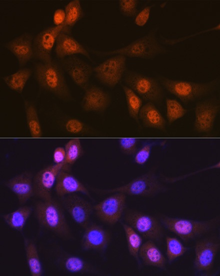
Immunofluorescence analysis of NIH-3T3 cells using Musashi-1 (MSI1) Rabbit mAb (CAB9122) at dilution of 1:100 (40x lens). Secondary antibody: Cy3-conjugated Goat anti-Rabbit IgG (H+L) (CABS007) at 1:500 dilution. Blue: DAPI for nuclear staining.


![Anti-Musashi 1 / MSI1 [ZN127] Monoclonal Antibody (AGMB06250)](https://cdn11.bigcommerce.com/s-h68l9z2lnx/images/stencil/590x590/products/277531/680949/anti-musashi-1-msi1-zn127-monoclonal-antibody-agmb06250__86639.1773043701.jpg?c=2 "Anti-Musashi 1 / MSI1 [ZN127] Monoclonal Antibody (AGMB06250)")

![Anti-Musashi 1 / MSI1 [R05-3A8] Monoclonal Antibody (AGMB02747)](https://cdn11.bigcommerce.com/s-h68l9z2lnx/images/stencil/590x590/products/274036/679384/anti-musashi-1-msi1-r05-3a8-monoclonal-antibody-agmb02747__77868.1773038775.jpg?c=2 "Anti-Musashi 1 / MSI1 [R05-3A8] Monoclonal Antibody (AGMB02747)")

