The STX1A Monoclonal Antibody (CAB19243) is a high-quality antibody developed for reliable detection and analysis of target proteins. This antibody, generated in rabbits, is highly specific to human samples and is suitable for use in various applications, including Western blotting and immunofluorescence.STX1A is a key player in neuronal communication, facilitating the release of neurotransmitters at synaptic terminals. Dysregulation of STX1A function has been implicated in neurological disorders such as epilepsy, Alzheimer's disease, and Parkinson's disease.
This antibody is validated for use in WB, IHC-P, ELISA, IF-P applications and has demonstrated reactivity against Human, Mouse, Rat samples.
Product Name:
STX1A Monoclonal Antibody
SKU:
CAB19243
Size:
20μL, 100μL
Reactivity:
Human, Mouse, Rat
Clone Number:
ARC2403
Conjugate:
Unconjugated
Immunogen:
Synthetic peptide. This information is considered to be commercially sensitive.
Recommended starting concentration is 1 μg/mL. Please optimize the concentration based on your specific assay requirements.
Synonyms:
STX1, HPC-1, P35-1, SYN1A, STX1A
Positive Sample:
C6, U-87 MG, Rat brain
Cellular Localization:
Cell Junction, Cell Membrane, Cytoplasmic Vesicle, Secreted, Single-Pass Type Iv Membrane Protein, Secretory Vesicle, Synapse, Synaptic Vesicle Membrane, Synaptosome.
Calculated MW:
33kDa
Observed MW:
33kDa
This gene encodes a member of the syntaxin superfamily. Syntaxins are nervous system-specific proteins implicated in the docking of synaptic vesicles with the presynaptic plasma membrane. Syntaxins possess a single C-terminal transmembrane domain, a SNARE [Soluble NSF (N-ethylmaleimide-sensitive fusion protein)-Attachment protein REceptor] domain (known as H3), and an N-terminal regulatory domain (Habc). Syntaxins bind synaptotagmin in a calcium-dependent fashion and interact with voltage dependent calcium and potassium channels via the C-terminal H3 domain. This gene product is a key molecule in ion channel regulation and synaptic exocytosis. Alternatively spliced transcript variants encoding different isoforms have been found for this gene.
Purification Method
Affinity purification
Gene ID
6804
Buffer Information
Store at -20℃. Avoid freeze / thaw cycles. Buffer: PBS containing 50% glycerol and 0.05% BSA, preserved with proclin300 or sodium azide, pH 7.3.
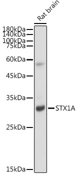
Western blot analysis of lysates from Rat brain, using STX1A Rabbit mAb (CAB19243) at 1:1000 dilution. Secondary antibody: HRP-conjugated Goat anti-Rabbit IgG (H+L) (CABS014) at 1:10000 dilution. Lysates/proteins: 25μg per lane. Blocking buffer: 3% nonfat dry milk in TBST. Detection: ECL Basic Kit (AbGn00020). Exposure time: 5s.
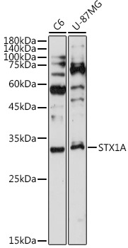
Western blot analysis of various lysates using STX1A Rabbit mAb (CAB19243) at 1:1000 dilution. Secondary antibody: HRP-conjugated Goat anti-Rabbit IgG (H+L) (CABS014) at 1:10000 dilution. Lysates/proteins: 25μg per lane. Blocking buffer: 3% nonfat dry milk in TBST. Detection: ECL Basic Kit (AbGn00020). Exposure time: 180s.
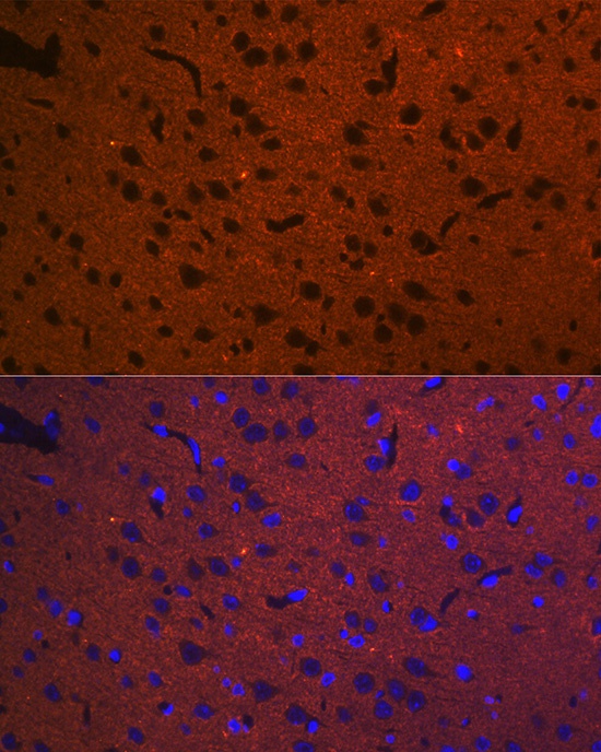
Immunofluorescence analysis of paraffin-embedded rat brain using STX1A Rabbit mAb (CAB19243) at dilution of 1:100 (40x lens). Secondary antibody: Cy3-conjugated Goat anti-Rabbit IgG (H+L) (CABS007) at 1:500 dilution. Blue: DAPI for nuclear staining.
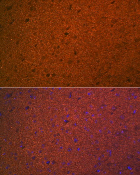
Immunofluorescence analysis of paraffin-embedded mouse brain using STX1A Rabbit mAb (CAB19243) at dilution of 1:100 (40x lens). Secondary antibody: Cy3-conjugated Goat anti-Rabbit IgG (H+L) (CABS007) at 1:500 dilution. Blue: DAPI for nuclear staining.