The TAX1BP3 Monoclonal Antibody (CAB19793) is a high-quality antibody developed for reliable detection and analysis of target proteins. This antibody, generated in rabbits, exhibits high specificity and sensitivity towards human samples, making it ideal for use in Western blotting and other immunological techniques. By targeting the Tax1BP3 protein, researchers can accurately detect and analyze its expression in different cell types, providing valuable insights into its role in autophagy-related pathways and cancer biology.Tax1BP3, also known as Tax1 binding protein 3, has been shown to play a critical role in the regulation of autophagy, a cellular process that helps maintain cellular homeostasis by degrading damaged organelles and proteins.
This antibody is validated for use in WB, ELISA applications and has demonstrated reactivity against Human, Mouse, Rat samples.
Product Name:
TAX1BP3 Monoclonal Antibody
SKU:
CAB19793
Size:
20μL, 100μL
Reactivity:
Human, Mouse, Rat
Clone Number:
ARC2319
Conjugate:
Unconjugated
Immunogen:
Synthetic peptide. This information is considered to be commercially sensitive.
Recommended starting concentration is 1 μg/mL. Please optimize the concentration based on your specific assay requirements.
Synonyms:
TIP1, TIP-1, TAX1BP3
Positive Sample:
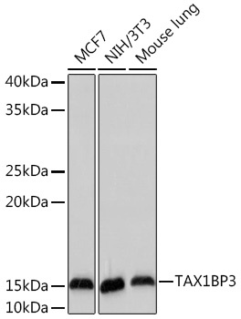
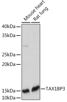
MCF7, NIH/3T3, Mouse lung, Mouse heart, Rat lung
Cellular Localization:
Actin Cytoskeleton, Cytoplasm, Cytosol, Extracellular Exosome, Fibrillar Center, Plasma Membrane.
Calculated MW:
14kDa
Observed MW:
14kDa
This gene encodes a small, highly conserved protein with a single PDZ domain. PDZ (PSD-95/Discs large/ZO-1 homologous) domains promote protein-protein interactions that affect cell signaling, adhesion, protein scaffolding, and receptor and ion transporter functions. The encoded protein interacts with a large number of target proteins that play roles in signaling pathways; for example, it interacts with Rho A and glutaminase L and also acts as a negative regulator of the Wnt/beta-catenin signaling pathway. This protein was first identified as binding to the T-cell leukaemia virus (HTLV1) Tax oncoprotein. Overexpression of this gene has been implicated in altered cancer cell adhesion, migration and metastasis. The encoded protein also modulates the localization and density of inwardly rectifying potassium channel 2.3 (Kir2.3). To date, this protein has been shown to play a role in cell proliferation, development, stress response, and polarization. Alternative splicing results in multiple transcript variants encoding distinct isoforms.
Purification Method
Affinity purification
Gene ID
30851
Buffer Information
Store at -20℃. Avoid freeze / thaw cycles. Buffer: PBS containing 50% glycerol and 0.05% BSA, preserved with proclin300 or sodium azide, pH 7.3.
Western blot analysis of various lysates using TAX1BP3 Rabbit mAb (CAB19793) at 1:1000 dilution. Secondary antibody: HRP-conjugated Goat anti-Rabbit IgG (H+L) (CABS014) at 1:10000 dilution. Lysates/proteins: 25μg per lane. Blocking buffer: 3% nonfat dry milk in TBST. Detection: ECL Basic Kit (AbGn00020). Exposure time: 180s.
Western blot analysis of various lysates using TAX1BP3 Rabbit mAb (CAB19793) at 1:1000 dilution. Secondary antibody: HRP-conjugated Goat anti-Rabbit IgG (H+L) (CABS014) at 1:10000 dilution. Lysates/proteins: 25μg per lane. Blocking buffer: 3% nonfat dry milk in TBST. Detection: ECL Enhanced Kit (AbGn00021). Exposure time: 180s.


![Anti-TAX1BP3 [R05-3N1] Monoclonal Antibody (AGMB02932)](https://cdn11.bigcommerce.com/s-h68l9z2lnx/images/stencil/590x590/products/274221/677109/anti-tax1bp3-r05-3n1-monoclonal-antibody-agmb02932__35767.1773031583.jpg?c=2 "Anti-TAX1BP3 [R05-3N1] Monoclonal Antibody (AGMB02932)")


