The CD86 Antibody (CAB1199) is a high-quality antibody developed for reliable detection and analysis of target proteins. This antibody, generated from rabbit hosts, exhibits high reactivity with human samples and is validated for use in Western blot applications. By specifically binding to the CD86 protein, this antibody enables reliable detection and analysis in a variety of cell types, making it an ideal choice for investigations in immunology and cancer research.CD86, also known as B7-2, plays a crucial role in T cell activation and immune response regulation. Its function as a co-stimulatory molecule allows CD86 to promote immune activation and cytokine production, leading to an enhanced immune response.
This antibody is validated for use in WB, IF/ICC, ELISA applications and has demonstrated reactivity against Human samples.
Product Name:
CD86 Antibody
SKU:
CAB1199
Size:
20μL, 100μL
Reactivity:
Human
Conjugate:
Unconjugated
Immunogen:
Recombinant protein (or fragment).This information is considered to be commercially sensitive.
Recommended starting concentration is 1 μg/mL. Please optimize the concentration based on your specific assay requirements.
Synonyms:
B70, B7-2, B7.2, LAB72, CD28LG2, CD86
Positive Sample:
Raji
Cellular Localization:
Cell Membrane, Single-Pass Type I Membrane Protein.
Calculated MW:
38kDa
Observed MW:
60-85kDa
This gene encodes a type I membrane protein that is a member of the immunoglobulin superfamily. This protein is expressed by antigen-presenting cells, and it is the ligand for two proteins at the cell surface of T cells, CD28 antigen and cytotoxic T-lymphocyte-associated protein 4. Binding of this protein with CD28 antigen is a costimulatory signal for activation of the T-cell. Binding of this protein with cytotoxic T-lymphocyte-associated protein 4 negatively regulates T-cell activation and diminishes the immune response. Alternative splicing results in several transcript variants encoding different isoforms.
Purification Method
Affinity purification
Gene ID
942
RRID
AB_2758917
Buffer Information
Store at -20℃. Avoid freeze / thaw cycles. Buffer: PBS containing 50% glycerol, preserved with proclin300 or sodium azide, pH 7.3.
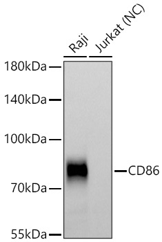
Western blot analysis of various lysates using CD86 Rabbit pAb (CAB1199) at 1:1000 dilution incubated overnight at 4℃. Secondary antibody: HRP-conjugated Goat anti-Rabbit IgG (H+L) (CABS014) at 1:10000 dilution. Lysates/proteins: 25 μg per lane. Blocking buffer: 3% nonfat dry milk in TBST. Detection: ECL Basic Kit (AbGn00020). Negative control (NC): Jurkat Exposure time: 90s.
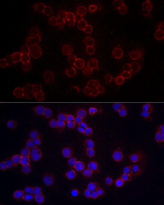
Immunofluorescence analysis of Raji cells using CD86 Rabbit pAb (CAB1199) at dilution of 1:200 (40x lens). Secondary antibody: Cy3-conjugated Goat anti-Rabbit IgG (H+L) (CABS007) at 1:500 dilution. Blue: DAPI for nuclear staining.