The PHD3 Antibody (CAB8001) is a high-quality antibody developed for reliable detection and analysis of target proteins. This polyclonal antibody, produced in rabbits, exhibits high reactivity with human samples and has been validated for use in Western blot applications.EGL-9 homolog 3 is a key player in the cellular response to changes in oxygen levels, making it crucial for understanding processes like angiogenesis, metabolism, and cell survival under hypoxic conditions. By targeting EGL-9 homolog 3 with this antibody, researchers can detect and analyze the protein in various cell types, providing insights into its role in oxygen sensing and HIF regulation.
This antibody is validated for use in WB, IHC-P, IF/ICC, ELISA applications and has demonstrated reactivity against Human, Mouse, Rat samples.
Product Name:
PHD3 Antibody
SKU:
CAB8001
Size:
20μL, 100μL
Reactivity:
Human, Mouse, Rat
Conjugate:
Unconjugated
Immunogen:
Synthetic peptide. This information is considered to be commercially sensitive.
Recommended starting concentration is 1 μg/mL. Please optimize the concentration based on your specific assay requirements.
Synonyms:
PHD3, HIFPH3, HIFP4H3
Positive Sample:
BxPC-3, Mouse brain, Mouse kidney, Rat stomach
Cellular Localization:
Cytoplasm, Nucleus.
Calculated MW:
27kDa
Observed MW:
32kDa
Enables peptidyl-proline 4-dioxygenase activity. Involved in several processes, including activation of cysteine-type endopeptidase activity involved in apoptotic process; peptidyl-proline hydroxylation to 4-hydroxy-L-proline; and response to hypoxia. Located in cytosol and nucleus. Implicated in renal cell carcinoma. Biomarker of clear cell renal cell carcinoma.
Purification Method
Affinity purification
Gene ID
112399
RRID
AB_2769278
Buffer Information
Store at -20℃. Avoid freeze / thaw cycles. Buffer: PBS containing 50% glycerol, preserved with proclin300 or sodium azide, pH 7.3.
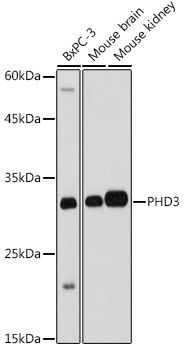
Western blot analysis of various lysates using PHD3 Rabbit pAb (CAB8001) at 1:1000 dilution. Secondary antibody: HRP-conjugated Goat anti-Rabbit IgG (H+L) (CABS014) at 1:10000 dilution. Lysates/proteins: 25μg per lane. Blocking buffer: 3% nonfat dry milk in TBST. Detection: ECL Basic Kit (AbGn00020). Exposure time: 1s.
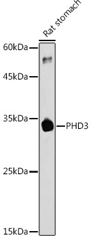
Western blot analysis of lysates from Rat stomach, using PHD3 Rabbit pAb (CAB8001) at 1:1000 dilution. Secondary antibody: HRP-conjugated Goat anti-Rabbit IgG (H+L) (CABS014) at 1:10000 dilution. Lysates/proteins: 25μg per lane. Blocking buffer: 3% nonfat dry milk in TBST. Detection: ECL Basic Kit (AbGn00020). Exposure time: 180s.
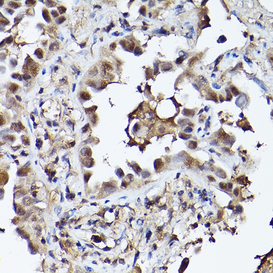
Immunohistochemistry analysis of paraffin-embedded Human lung cancer using PHD3 Rabbit pAb (CAB8001) at dilution of 1:100 (40x lens). High pressure antigen retrieval performed with 0.01M Citrate buffer (pH 6.0) prior to IHC staining.
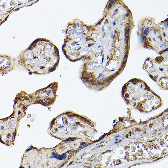
Immunohistochemistry analysis of paraffin-embedded Human placenta using PHD3 Rabbit pAb (CAB8001) at dilution of 1:100 (40x lens). High pressure antigen retrieval performed with 0.01M Citrate buffer (pH 6.0) prior to IHC staining.