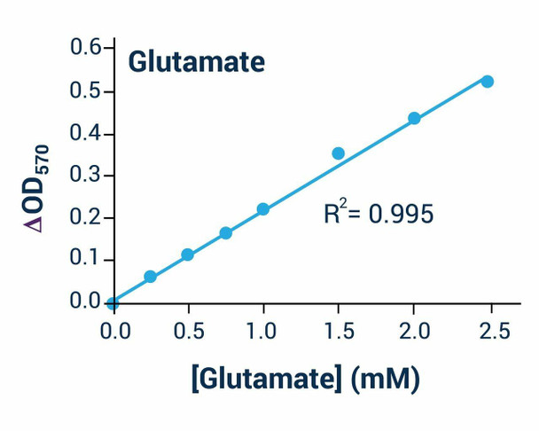
Description
Glutamate Assay Kit (Colorimetric) (BA0114) (BA0114)
The Glutamate Assay Kit (Colorimetric) (SKU: BA0114) provides a simple, direct and automation-ready colorimetric method for measuring glutamate. Glutamate is an important metabolite and a crucial mammalian neurotransmitter implicated in a range of neurological and psychiatric disorders, as well as a widely used flavour enhancer. The assay is based on glutamate dehydrogenase-catalysed oxidation of glutamate, in which the NADH formed reduces an MTT reagent. The intensity of the product colour, measured at 565 nm, is proportional to the glutamate concentration in the sample. The room-temperature procedure requires no 37C heater and is readily automated for high-throughput work.
| Product Name: | Glutamate Assay Kit (Colorimetric) (BA0114) |
| SKU: | BA0114 |
| Detection Method: | Colorimetric (565 nm) |
| Detection Range: | 0.05 - 2.5 mM glutamate (detection limit 50 uM) |
| Sample Type: | Serum, plasma, tissue extracts and food extract samples |
| Species Reactivity: | All |
| Assay Time: | 30 minutes |
| Kit Size: | 100 Assays |
| Equipment Required: | Microplate reader |
| Storage: | -20C |
| Shelf Life: | 6 months after receipt |
| Shipping: | Gel Pack |
A colorimetric assay for glutamate in biological and food samples using a single working reagent read at time zero and 30 minutes. Serum and tissue extract samples require a sample blank.
- Sensitive and accurate with a detection limit of 50 uM and linearity up to 2.5 mM glutamate
- Convenient single working reagent read at time zero and 30 minutes, no 37C heater required
- High-throughput and readily automated
- Direct assay of glutamate in serum, plasma, tissue extracts and food extract samples
- Studying the effects of drugs on glutamate levels
Note: The below protocol is a sample protocol. Protocols are specific to each batch/lot. For the correct instructions please follow the protocol included in your kit.
| Step | Procedure |
| 1 | Calibration Curve: prepare 600 uL of 2.5 mM Glutamate Premix by mixing 15 uL of 100 mM Standard with 585 uL distilled water, then dilute per the table. Transfer 20 uL of each standard into a clear-bottom 96-well plate. |
| 2 | Samples: add 20 uL of each sample to separate wells. Serum and tissue extract samples require a sample blank. |
| 3 | Reagent Preparation: spin enzyme tubes briefly. For each well mix 60 uL Assay Buffer, 1 uL Enzyme A, 1 uL Enzyme B, 5 uL NAD and 14 uL MTT. For sample blanks omit Enzyme A. |
| 4 | Reaction: add 80 uL of the appropriate reagent per well quickly and tap to mix. |
| 5 | Read optical density (OD0) at 565 nm (520-600 nm) at time zero and OD30 after 30 minutes at room temperature. |
Subtract OD0 from OD30 for standards and samples, then subtract the water delta-OD (standard 8) to obtain delta-delta-OD values (for sample blanks subtract the blank delta-OD). Plot the standard delta-delta-OD values and convert sample readings to concentration: [Glutamate] = delta-delta-ODSAMPLE / Slope (mM). If a reading exceeds that of the 2.5 mM standard, dilute in distilled water, repeat and multiply by the dilution factor. 1 mM glutamate equals 14.6 mg/dL.
| Component | Quantity | Storage |
| Assay Buffer | 10 mL | -20C |
| NAD Solution | 1 mL | -20C |
| MTT Solution | 1.5 mL | -20C |
| Enzyme A | 120 uL | -20C |
| Enzyme B | 120 uL | -20C |
| Standard (100 mM Glutamate) | 1 mL | -20C |