The KHSRP Monoclonal Antibody (CAB9075) is a high-quality antibody developed for reliable detection and analysis of target proteins. This antibody, generated in rabbits, is highly specific for human samples and is validated for use in applications such as Western blotting. It specifically binds to the KHSRP protein, allowing for its detection and analysis in a variety of cell types.KHSRP, also known as KH-type splicing regulatory protein, plays a crucial role in regulating the expression of genes involved in processes such as RNA processing, stability, and translation.
This antibody is validated for use in WB, IHC-P, IP, ELISA applications and has demonstrated reactivity against Human, Mouse, Rat samples.
Product Name:
KHSRP Monoclonal Antibody
SKU:
CAB9075
Size:
20μL, 100μL
Reactivity:
Human, Mouse, Rat
Clone Number:
ARC1402
Conjugate:
Unconjugated
Immunogen:
Recombinant protein (or fragment).This information is considered to be commercially sensitive.
0.5μg-4μg antibody for 200μg-400μg extracts of whole cells
ELISA
Recommended starting concentration is 1 μg/mL. Please optimize the concentration based on your specific assay requirements.
Synonyms:
p75, FBP2, KSRP, FUBP2, KHSRP
Positive Sample:
Hep G2, A549, C2C12
Cellular Localization:
Cytoplasm, Nucleus.
Calculated MW:
73kDa
Observed MW:
82kDa
The KHSRP gene encodes a multifunctional RNA-binding protein implicated in a variety of cellular processes, including transcription, alternative pre-mRNA splicing, and mRNA localization (Min et al., 1997 [PubMed 9136930]; Gherzi et al., 2004 [PubMed 15175153]).
Purification Method
Affinity purification
Gene ID
8570
RRID
AB_2863652
Buffer Information
Store at -20℃. Avoid freeze / thaw cycles. Buffer: PBS containing 50% glycerol and 0.05% BSA, preserved with proclin300 or sodium azide, pH 7.3.
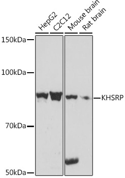
Western blot analysis of various lysates using KHSRP Rabbit mAb (CAB9075) at 1:1000 dilution. Secondary antibody: HRP-conjugated Goat anti-Rabbit IgG (H+L) (CABS014) at 1:10000 dilution. Lysates/proteins: 25μg per lane. Blocking buffer: 3% nonfat dry milk in TBST. Detection: ECL Basic Kit (AbGn00020). Exposure time: 10s.
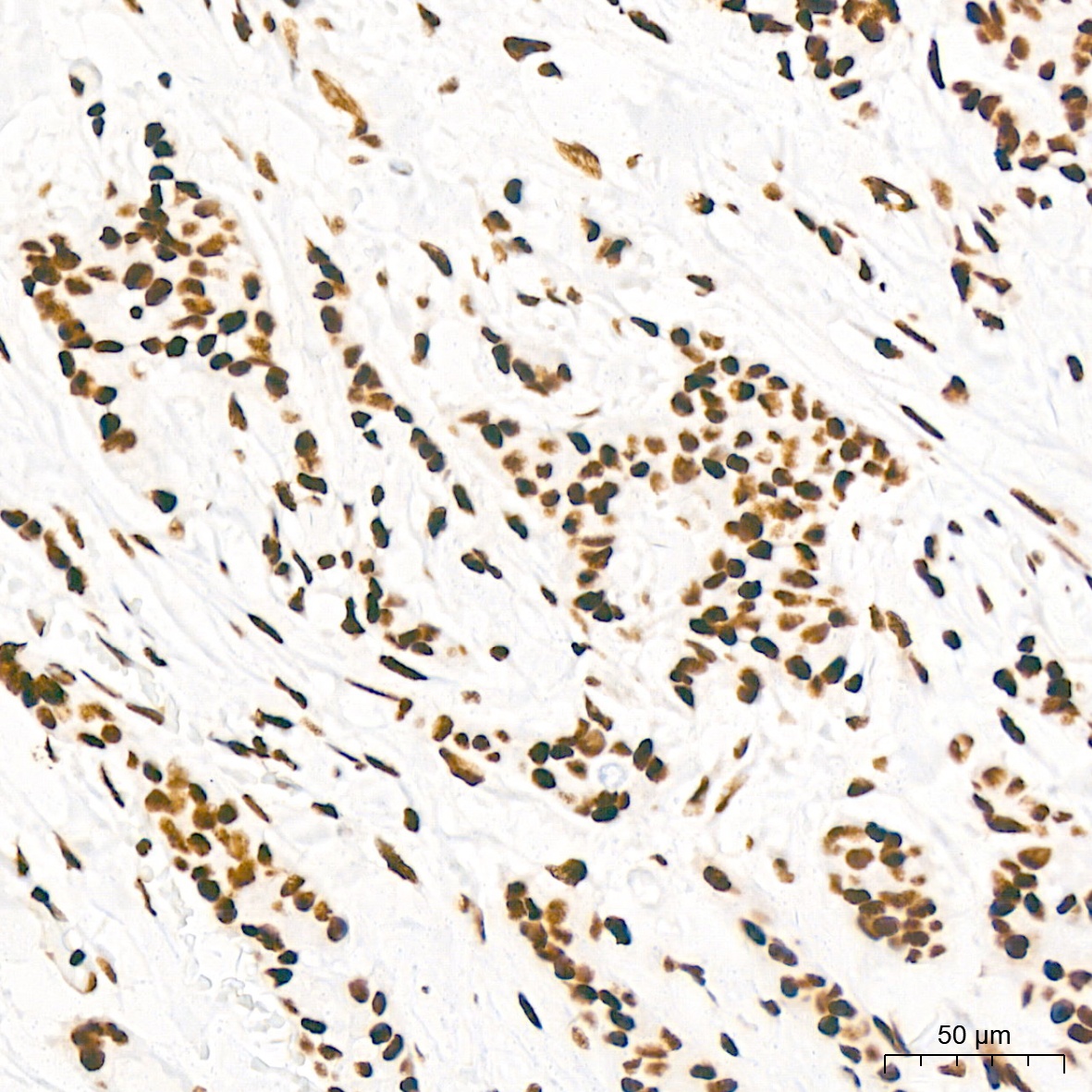
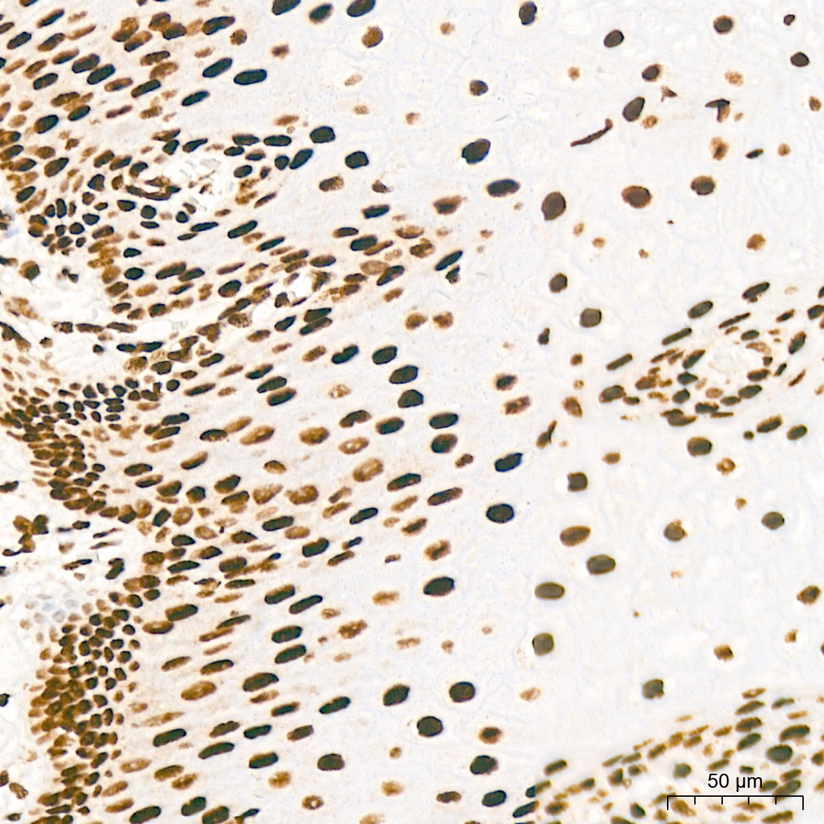
Immunohistochemistry analysis of paraffin-embedded Human breast cancer tissue using KHSRP Rabbit mAb (CAB9075) at a dilution of 1:200 (40x lens). High pressure antigen retrieval performed with 0.01M Citrate buffer (pH 6.0) prior to IHC staining.
Immunohistochemistry analysis of paraffin-embedded Human esophagus tissue using KHSRP Rabbit mAb (CAB9075) at a dilution of 1:200 (40x lens). High pressure antigen retrieval performed with 0.01M Citrate buffer (pH 6.0) prior to IHC staining.
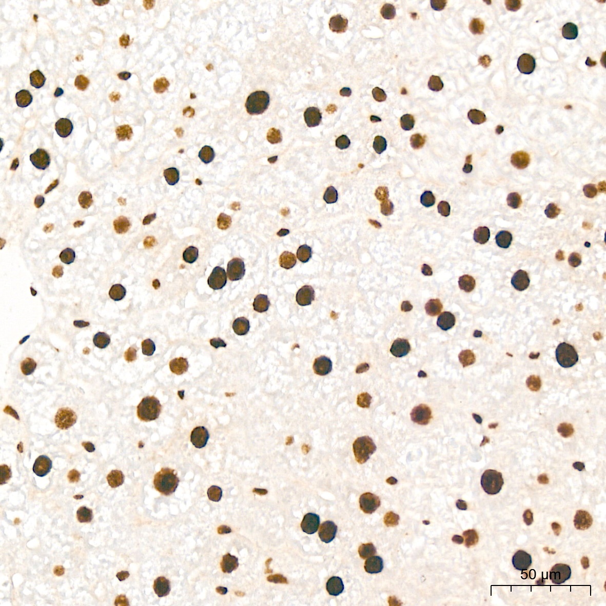
Immunohistochemistry analysis of paraffin-embedded Human thyroid tissue using KHSRP Rabbit mAb (CAB9075) at a dilution of 1:200 (40x lens). High pressure antigen retrieval performed with 0.01M Citrate buffer (pH 6.0) prior to IHC staining.
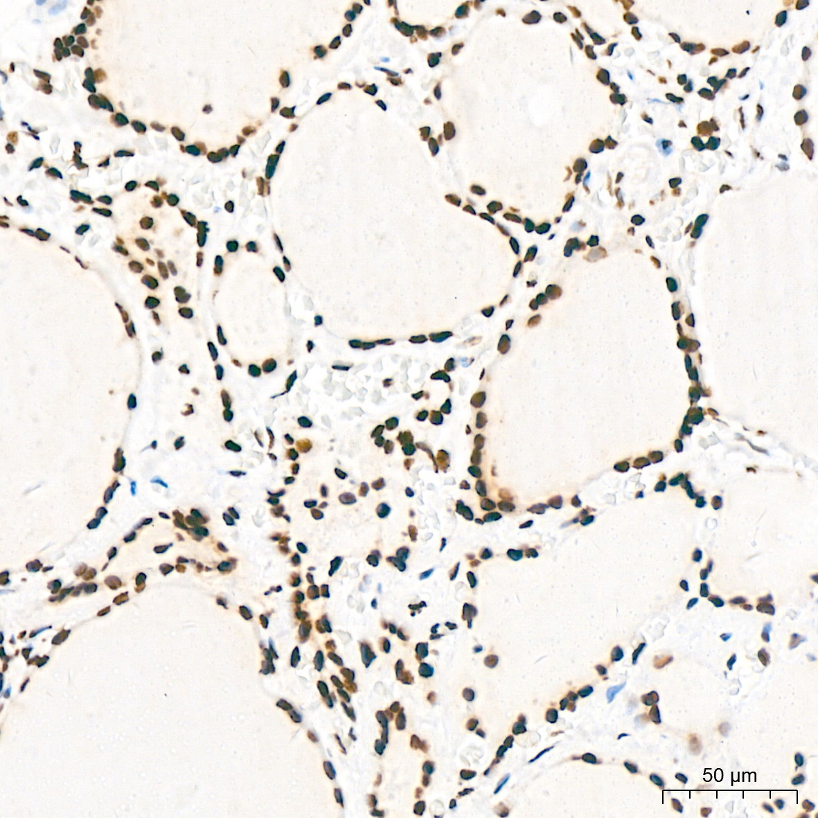
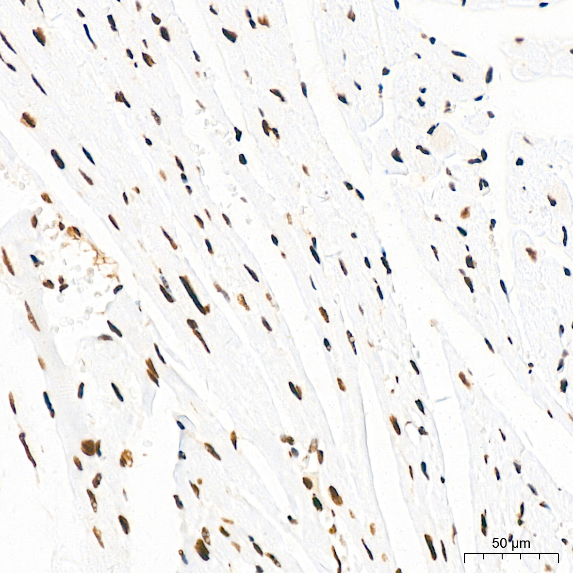
Immunohistochemistry analysis of paraffin-embedded Mouse heart tissue using KHSRP Rabbit mAb (CAB9075) at a dilution of 1:200 (40x lens). High pressure antigen retrieval performed with 0.01M Citrate buffer (pH 6.0) prior to IHC staining.
Immunohistochemistry analysis of paraffin-embedded Mouse liver tissue using KHSRP Rabbit mAb (CAB9075) at a dilution of 1:200 (40x lens). High pressure antigen retrieval performed with 0.01M Citrate buffer (pH 6.0) prior to IHC staining.
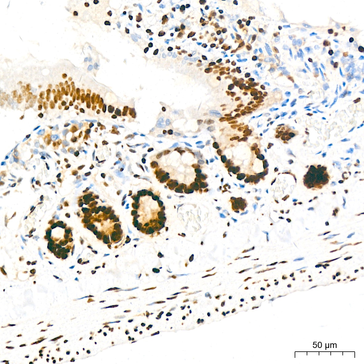
Immunohistochemistry analysis of paraffin-embedded Rat colon tissue using KHSRP Rabbit mAb (CAB9075) at a dilution of 1:200 (40x lens). High pressure antigen retrieval performed with 0.01M Citrate buffer (pH 6.0) prior to IHC staining.
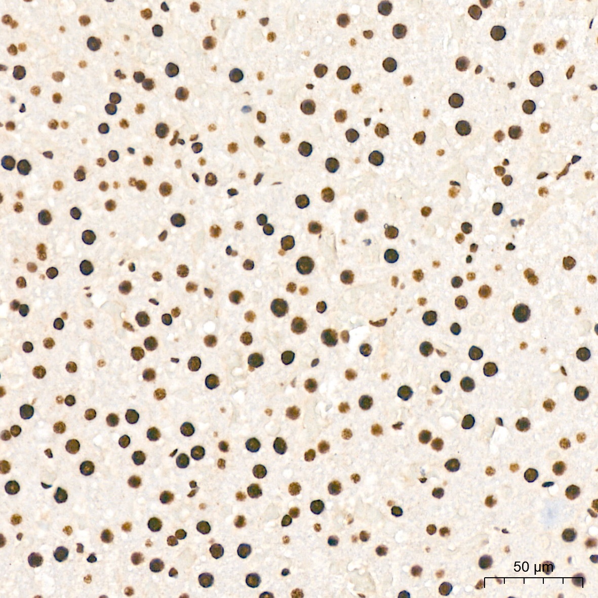
Immunohistochemistry analysis of paraffin-embedded Rat liver tissue using KHSRP Rabbit mAb (CAB9075) at a dilution of 1:200 (40x lens). High pressure antigen retrieval performed with 0.01M Citrate buffer (pH 6.0) prior to IHC staining.


![Anti-KHSRP [R09-9E7] Monoclonal Antibody (AGMB00443)](https://cdn11.bigcommerce.com/s-h68l9z2lnx/images/stencil/590x590/products/271732/691013/anti-khsrp-r09-9e7-monoclonal-antibody-agmb00443__72493.1774501647.jpg?c=2 "Anti-KHSRP [R09-9E7] Monoclonal Antibody (AGMB00443)")


