The [KO Validated] Lamin A/C Monoclonal Antibody (CAB19524) is a high-quality antibody developed for reliable detection and analysis of target proteins. This antibody, produced in rabbits, is highly specific to human samples and has been rigorously validated for use in immunohistochemistry and immunofluorescence applications.Lamin A/C is essential for proper cell function and integrity, with mutations in the LMNA gene associated with a range of diseases including muscular dystrophies and premature aging syndromes.
This antibody is validated for use in WB, IHC-P, IF/ICC, IP, ELISA applications and has demonstrated reactivity against Human, Mouse, Rat samples.
Product Name:
[KO Validated] Lamin A/C Monoclonal Antibody
SKU:
CAB19524
Size:
20μL, 100μL
Reactivity:
Human, Mouse, Rat
Clone Number:
ARC5001-08
Conjugate:
Unconjugated
Immunogen:
Recombinant protein (or fragment).This information is considered to be commercially sensitive.
The protein encoded by this gene is part of the nuclear lamina, a two-dimensional matrix of proteins located next to the inner nuclear membrane. The lamin family of proteins make up the matrix and are highly conserved in evolution. During mitosis, the lamina matrix is reversibly disassembled as the lamin proteins are phosphorylated. Lamin proteins are thought to be involved in nuclear stability, chromatin structure and gene expression. Vertebrate lamins consist of two types, A and B. Alternative splicing results in multiple transcript variants. Mutations in this gene lead to several diseases: Emery-Dreifuss muscular dystrophy, familial partial lipodystrophy, limb girdle muscular dystrophy, dilated cardiomyopathy, Charcot-Marie-Tooth disease, and Hutchinson-Gilford progeria syndrome.
Purification Method
Affinity purification
Gene ID
4000
RRID
AB_2862647
Buffer Information
Store at -20℃. Avoid freeze / thaw cycles. Buffer: PBS containing 50% glycerol and 0.05% BSA, preserved with proclin300 or sodium azide, pH 7.3.
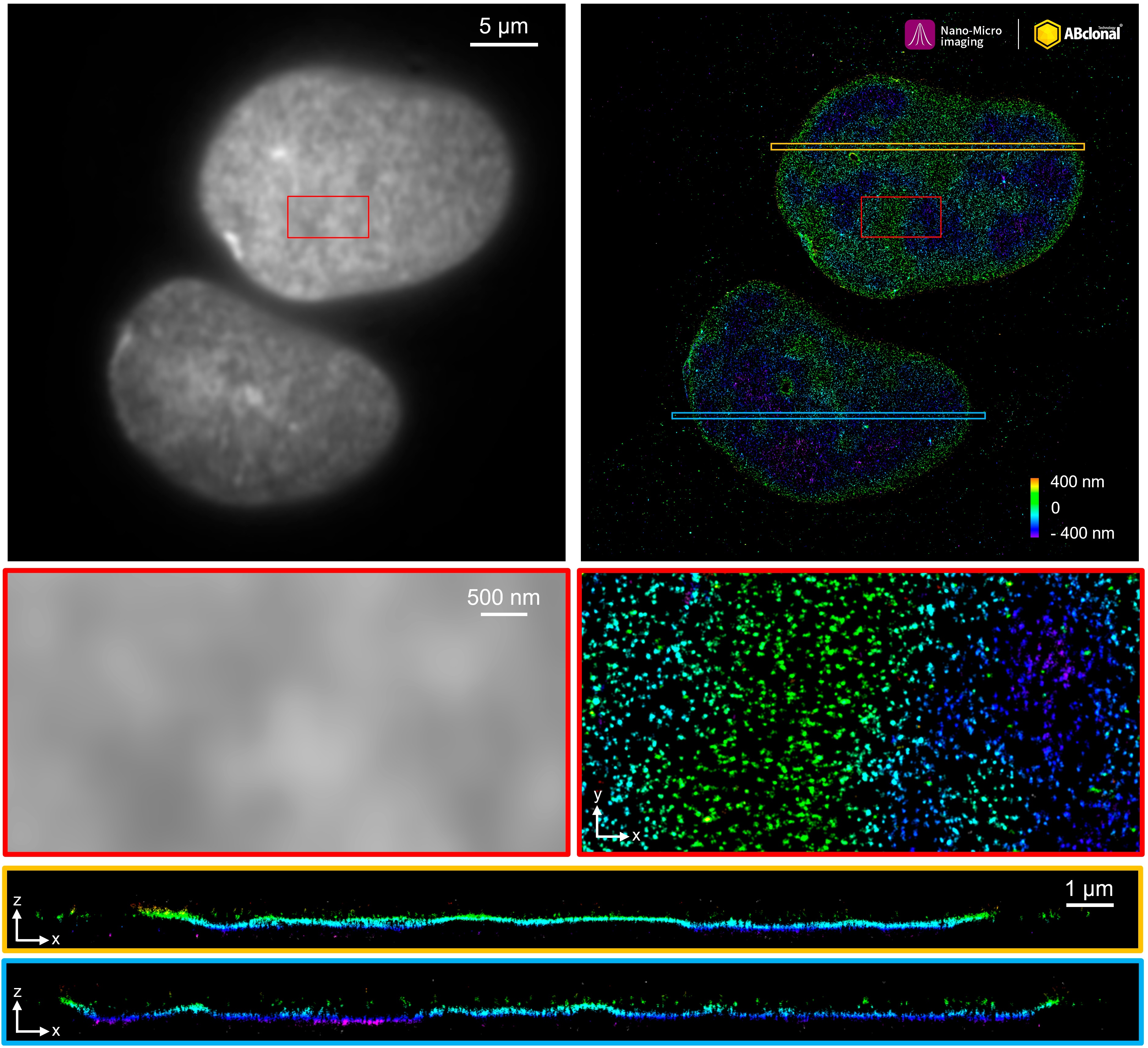
The STORM super-resolution (SR) imaging of U-2 OS cells using [KO Validated] Lamin A/C Rabbit mAb (CAB19524, ABclonal) at dilution of 1:200 with 3% paraformaldehyde (PFA) +0.1% glutaraldehyde (GA) fixation. The immunostaining was performed by Full Automatic Immunofluorescence Workflow System (Workflow Ultra300, Nano-Micro imaging, China). Image was performed with Single-Molecule Localization Super-Resolution Microscopy (STORM Ultra300, Nano-Micro imaging, China). We acknowledge Nano-Micro imaging Biotechnology Co., Ltd. in SR image processing and kindly providing this image.
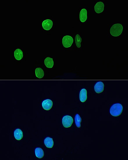
Immunofluorescence analysis of H9C2 cells using [KO Validated] Lamin A/C Rabbit mAb (CAB19524) at dilution of 1:200. Blue: DAPI for nuclear staining.
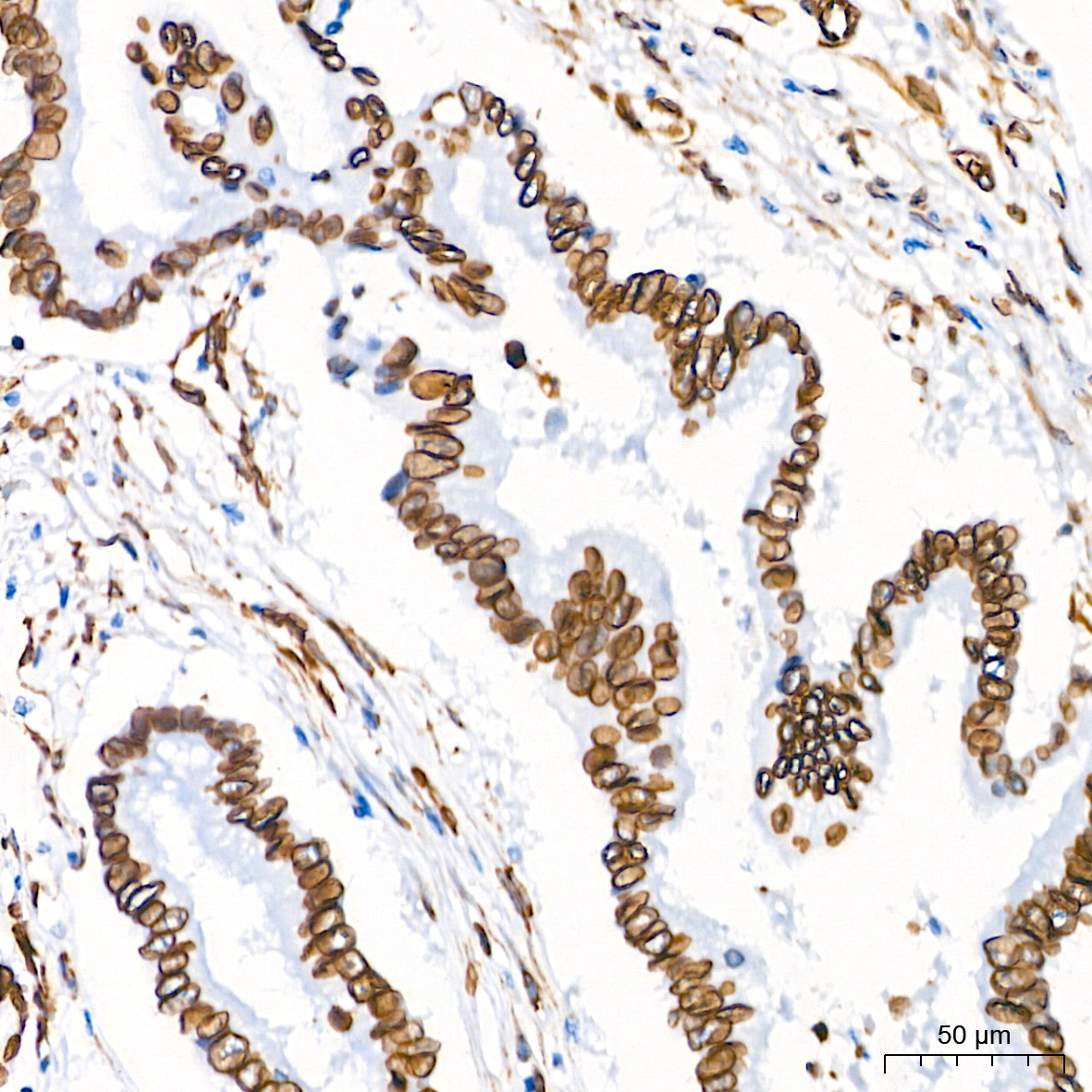
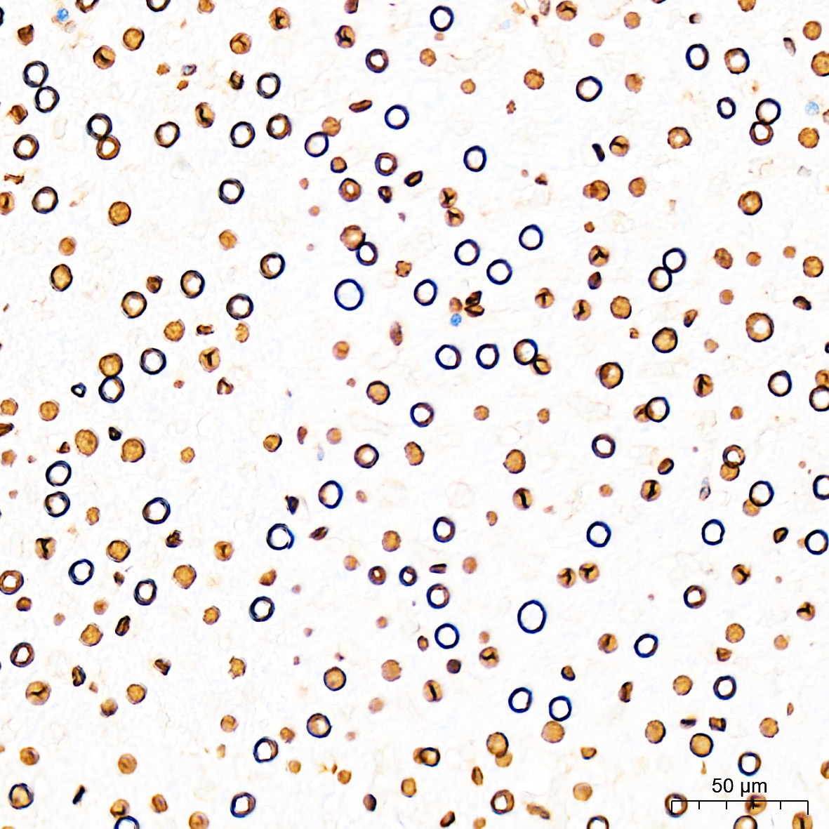
Immunohistochemistry analysis of paraffin-embedded Human colon carcinoma tissue using [KO Validated] Lamin A/C Rabbit mAb (CAB19524) at a dilution of 1:1300 (40x lens). High pressure antigen retrieval performed with 0.01M Citrate Buffer (pH 6.0) prior to IHC staining.
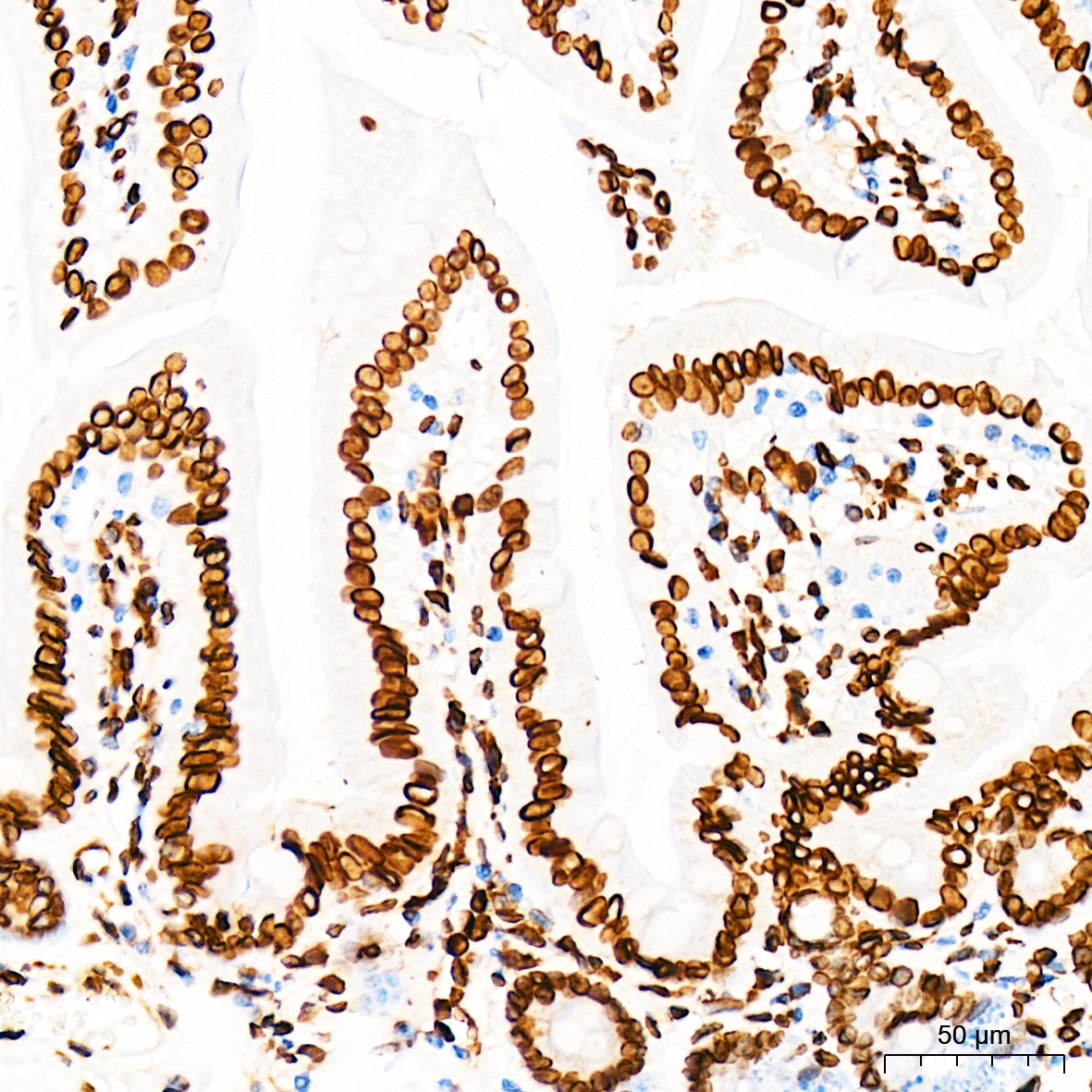
Immunohistochemistry analysis of paraffin-embedded Mouse intestin tissue using [KO Validated] Lamin A/C Rabbit mAb (CAB19524) at a dilution of 1:1300 (40x lens). High pressure antigen retrieval performed with 0.01M Citrate Buffer (pH 6.0) prior to IHC staining.
Immunohistochemistry analysis of paraffin-embedded Mouse liver tissue using [KO Validated] Lamin A/C Rabbit mAb (CAB19524) at a dilution of 1:1300 (40x lens). High pressure antigen retrieval performed with 0.01M Citrate Buffer (pH 6.0) prior to IHC staining.
Immunohistochemistry analysis of paraffin-embedded Rat liver tissue using [KO Validated] Lamin A/C Rabbit mAb (CAB19524) at a dilution of 1:1300 (40x lens). High pressure antigen retrieval performed with 0.01M Citrate Buffer (pH 6.0) prior to IHC staining.
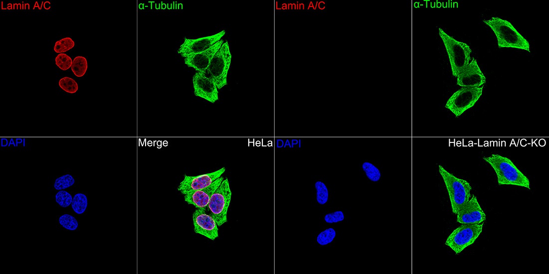
Confocal imaging of HeLa cells and Lamin A/C knockout(KO) HeLa cells using [KO Validated] Lamin A/C Rabbit mAb (CAB19524, dilution 1:200) followed by a further incubation with Cy3-conjugated Goat anti-Rabbit IgG (H+L) (CABS007, dilution 1:500) (Red). The cells were counterstained with α-Tubulin Mouse mAb (AC012, dilution 1:400) followed by incubation with ABflo® 488-conjugated Goat Anti-Mouse IgG (H+L) Ab (CABS076, dilution 1:500) (Green). DAPI was used for nuclear staining (Blue). Objective: 100x.
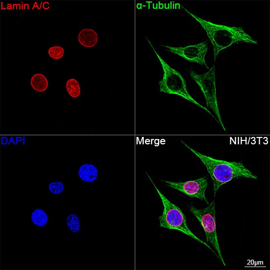
Confocal imaging of NIH/3T3 using [KO Validated] Lamin A/C Rabbit mAb (CAB19524, dilution 1:200) followed by a further incubation with Cy3-conjugated Goat anti-Rabbit IgG (H+L) (CABS007, dilution 1:500) (Red). The cells were counterstained with α-Tubulin Mouse mAb (AC012, dilution 1:400) followed by incubation with ABflo® 488-conjugated Goat Anti-Mouse IgG (H+L) Ab (CABS076, dilution 1:500) (Green). DAPI was used for nuclear staining (Blue). Objective: 100x.
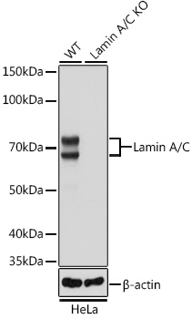
Western blot analysis of lysates from wild type (WT) and Lamin A/C knockout (KO) HeLa cells using [KO Validated] Lamin A/C Rabbit mAb (CAB19524) at 1:50000 dilution incubated overnight at 4℃. Secondary antibody: HRP-conjugated Goat anti-Rabbit IgG (H+L) (CABS014) at 1:10000 dilution. Lysates/proteins: 25μg per lane. Blocking buffer: 3% nonfat dry milk in TBST. Detection: ECL Basic Kit (AbGn00020). Exposure time: 1s.
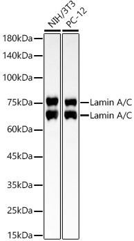
Western blot analysis of various lysates using [KO Validated] Lamin A/C Rabbit mAb (CAB19524) at 1:300000 dilution incubated overnight at 4℃. Secondary antibody: HRP-conjugated Goat anti-Rabbit IgG (H+L) (CABS014) at 1:10000 dilution. Lysates/proteins: 25μg per lane. Blocking buffer: 3% nonfat dry milk in TBST. Detection: ECL Basic Kit (AbGn00020). Exposure time: 30s.
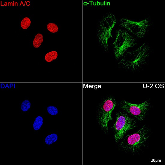
Confocal imaging of U-2 OS cells using [KO Validated] Lamin A/C Rabbit mAb (CAB19524, dilution 1:200) followed by a further incubation with Cy3 Goat Anti-Rabbit IgG (H+L) (CABS007, dilution 1:500) (Red). The cells were counterstained with α-Tubulin Mouse mAb (AC012, dilution 1:400) followed by incubation with ABflo® 488-conjugated Goat Anti-Mouse IgG (H+L) Ab (CABS076, dilution 1:500) (Green). DAPI was used for nuclear staining (Blue). Objective: 100x.