Description
L- Lactate Assay Kit (Colorimetric) (BA0091) (BA0091)
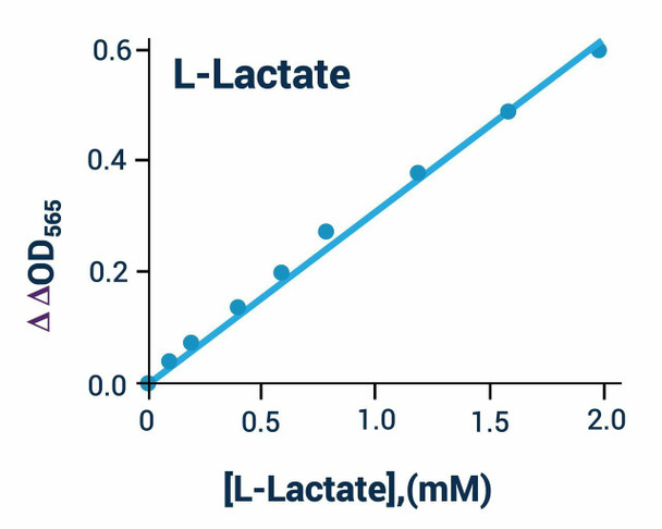
The L-Lactate Assay Kit (Colorimetric) (SKU: BA0091) provides a simple, direct and automation-ready procedure for measuring L-lactate concentration. Lactate is generated by lactate dehydrogenase under hypoxic or anaerobic conditions, so monitoring lactate levels is a good indicator of the balance between tissue oxygen demand and utilisation and is useful in cellular and animal physiology studies. This assay is based on lactate dehydrogenase-catalysed oxidation of lactate, in which the formed NADH reduces a formazan (MTT) reagent. The intensity of the product colour, measured at 565 nm, is proportional to the lactate concentration in the sample.
| Product Name: | L- Lactate Assay Kit (Colorimetric) (BA0091) |
| SKU: | BA0091 |
| Detection Method: | Colorimetric |
| Detection Range: | 0.05 to 2 mM (water); 0.1 to 1 mM (phenol red media) |
| Sample Type: | Serum, plasma and cell media |
| Species Reactivity: | All |
| Assay Time: | 20 minutes at room temperature |
| Kit Size: | 100 Assays |
| Equipment Required: | Microplate reader |
| Storage: | -20C |
| Shelf Life: | 6 months after receipt |
| Shipping: | Gel Pack |
Quantitative colorimetric determination of L-lactate at 565 nm. Lactate dehydrogenase oxidises lactate, generating NADH that reduces an MTT formazan reagent; the colour is proportional to lactate concentration.
- Sensitive and accurate; detection limit 0.05 mM and linearity up to 2 mM L-lactate
- Detection limit 0.1 mM and linearity up to 1 mM for phenol red cell culture samples
- Convenient single-working-reagent, room-temperature assay with no 37C heater required
- High-throughput; readily automated for thousands of samples per day
- Direct assays of lactate in serum, plasma and cell media samples
Note: The below protocol is a sample protocol. Protocols are specific to each batch/lot. For the correct instructions please follow the protocol included in your kit.
| Step | Procedure |
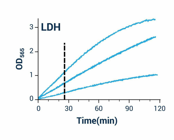
| 1 | This assay is a kinetic reaction; add working reagent quickly and mix briefly but thoroughly, using a multi-channel pipettor. |
| 2 | Standard curve. Prepare 1000 uL of 2.0 mM L-lactate premix by mixing 100 uL of 20 mM standard and 900 uL distilled water. For phenol red cell culture samples, prepare 1000 uL of 1.0 mM premix by mixing 50 uL of 20 mM standard and 950 uL serum-free culture medium. Dilute as shown in the dilution table and transfer 20 uL standards into wells of a clear bottom 96-well plate. |
| 3 | Samples. Add 20 uL sample per well in separate wells. For samples with potential endogenous enzyme activity (serum, plasma, tissue extracts), run two reactions: one with added Enzyme A and one No Enzyme A control. Dilute serum and plasma at least 2-fold with distilled water prior to assay. |
| 4 | Reagent preparation. Spin the enzyme tubes briefly. For each sample and standard well, prepare working reagent by mixing 60 uL Assay Buffer, 1 uL Enzyme A, 1 uL Enzyme B, 10 uL NAD and 14 uL MTT. For the No Enzyme A control, omit Enzyme A. |
| 5 | Reaction. Add 80 uL working reagent per reaction well quickly and tap to mix briefly and thoroughly. |
| 6 | Read optical density (OD0) for time zero at 565 nm (520-600 nm) and OD20 after a 20 minute incubation at room temperature. |
Subtract OD0 from OD20 for the standard and sample wells and use the dOD values to determine sample L-lactate concentration from the standard curve. For samples requiring a No Enzyme A control, subtract the dOD (No Enzyme A) value from the dOD (Sample) and use this ddOD value with the standard curve. If the sample OD exceeds that of the 2 mM standard, dilute the sample in water, repeat and multiply by the dilution factor.
| Component | Quantity | Storage |
| Assay Buffer | 10 mL | -20C |
| NAD Solution | 1 mL | -20C |
| Enzyme A | 120 uL | -20C |
| Enzyme B | 120 uL | -20C |
| MTT Solution | 1.5 mL | -20C |
| Standard (20 mM L-Lactate) | 1.0 mL | -20C |