The Cation-independent M6PR (IGF2R) Monoclonal Antibody (CAB3762) is a high-quality antibody developed for reliable detection and analysis of target proteins. This antibody, produced from rabbit cells, exhibits high specificity and sensitivity in detecting M6PR in human samples, making it a reliable choice for Western blot applications.The M6PR protein plays a crucial role in cellular processes such as lysosomal enzyme trafficking and autophagy, making it a key player in maintaining cellular homeostasis. Dysregulation of M6PR has been implicated in various diseases, including lysosomal storage disorders and cancer.
This antibody is validated for use in WB, IF/ICC, ELISA applications and has demonstrated reactivity against Human, Mouse, Rat samples.
Lysosome Membrane, Single-Pass Type I Membrane Protein.
Calculated MW:
274kDa
Observed MW:
274kDa
This gene encodes a receptor for both insulin-like growth factor 2 and mannose 6-phosphate. The binding sites for each ligand are located on different segments of the protein. This receptor has various functions, including in the intracellular trafficking of lysosomal enzymes, the activation of transforming growth factor beta, and the degradation of insulin-like growth factor 2. Mutation or loss of heterozygosity of this gene has been association with risk of hepatocellular carcinoma. The orthologous mouse gene is imprinted and shows exclusive expression from the maternal allele; however, imprinting of the human gene may be polymorphic, as only a minority of individuals showed biased expression from the maternal allele (PMID:8267611).
Purification Method
Affinity purification
Gene ID
3482
RRID
AB_2863138
Buffer Information
Store at -20℃. Avoid freeze / thaw cycles. Buffer: PBS containing 50% glycerol and 0.05% BSA, preserved with proclin300 or sodium azide, pH 7.3.
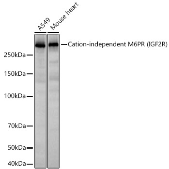
Western blot analysis of various lysates using Cation-independent M6PR (IGF2R) Rabbit mAb (CAB3762) at 1:3000 dilution incubated at room temperature for 1.5 hours. Secondary antibody: HRP-conjugated Goat anti-Rabbit IgG (H+L) (CABS014) at 1:10000 dilution. Lysates/proteins: 25 μg per lane. Blocking buffer: 3% nonfat dry milk in TBST. Detection: ECL Basic Kit (AbGn00020). Exposure time: 20 s.
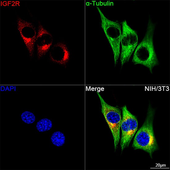
Confocal imaging of NIH/3T3 cells using Cation-independent M6PR (IGF2R) Rabbit mAb (CAB3762,dilution 1:100)(Red). The cells were counterstained with α-Tubulin Mouse mAb (AC012,dilution 1:400) (Green). DAPI was used for nuclear staining (blue). Objective: 100x.


 ELISA Kit (HUEB1839)")
 ELISA Kit (BOEB0814)")
 ELISA Kit (MOEB1572)")
![Anti-M6PR [R09-8E3] Monoclonal Antibody (AGMB03242)](https://cdn11.bigcommerce.com/s-h68l9z2lnx/images/stencil/590x590/products/274531/680465/anti-m6pr-r09-8e3-monoclonal-antibody-agmb03242__70291.1773042152.jpg?c=2 "Anti-M6PR [R09-8E3] Monoclonal Antibody (AGMB03242)")