The MCCC2 Antibody (CAB7990) is a high-quality antibody developed for reliable detection and analysis of target proteins. This antibody, produced in rabbits, exhibits high reactivity with human samples and has been validated for use in Western blot applications. By binding specifically to the MCCC2 protein, this antibody enables accurate detection and analysis in a variety of cell types, making it ideal for studies in metabolic disease, mitochondrial dysfunction, and related research areas.MCCC2, also known as methylcrotonoyl-CoA carboxylase 2, plays a crucial role in the breakdown of amino acids and fatty acids for energy production in mitochondria.
This antibody is validated for use in WB, IHC-P, ELISA applications and has demonstrated reactivity against Human, Mouse, Rat samples.
Product Name:
MCCC2 Antibody
SKU:
CAB7990
Size:
20μL, 100μL
Reactivity:
Human, Mouse, Rat
Conjugate:
Unconjugated
Immunogen:
Recombinant protein (or fragment).This information is considered to be commercially sensitive.
Recommended starting concentration is 1 μg/mL. Please optimize the concentration based on your specific assay requirements.
Synonyms:
MCCB, MCCCbeta, MCCC2
Positive Sample:
MCF7
Cellular Localization:
Mitochondrion Matrix.
Calculated MW:
61kDa
Observed MW:
61kDa
This gene encodes the small subunit of 3-methylcrotonyl-CoA carboxylase. This enzyme functions as a heterodimer and catalyzes the carboxylation of 3-methylcrotonyl-CoA to form 3-methylglutaconyl-CoA. Mutations in this gene are associated with 3-Methylcrotonylglycinuria, an autosomal recessive disorder of leucine catabolism. Alternative splicing results in multiple transcript variants encoding distinct isoforms.
Purification Method
Affinity purification
Gene ID
64087
RRID
AB_2770315
Buffer Information
Store at -20℃. Avoid freeze / thaw cycles. Buffer: PBS containing 50% glycerol, preserved with proclin300 or sodium azide, pH 7.3.
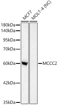
Western blot analysis of various lysates using MCCC2 Rabbit pAb (CAB7990) at 1:2000 dilution. Secondary antibody: HRP-conjugated Goat anti-Rabbit IgG (H+L) (CABS014) at 1:10000 dilution. Lysates/proteins: 25 μg per lane. Blocking buffer: 3% nonfat dry milk in TBST. Detection: ECL Basic Kit (AbGn00020). Negative control (NC): MOLT-4. Exposure time: 30s.
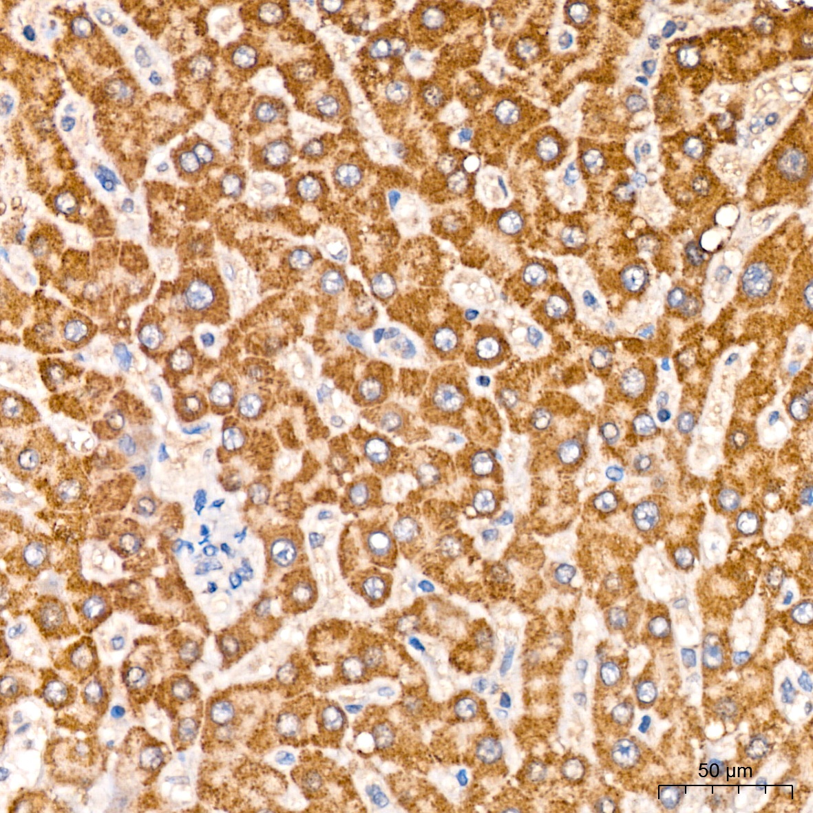
Immunohistochemistry analysis of paraffin-embedded Human liver tissue using MCCC2 Rabbit pAb (CAB7990) at a dilution of 1:200 (40x lens). High pressure antigen retrieval was performed with 0.01 M citrate buffer (pH 6.0) prior to IHC staining.
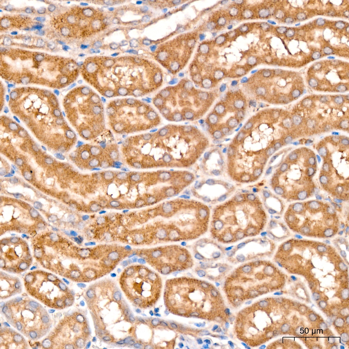
Immunohistochemistry analysis of paraffin-embedded Rat kidney tissue using MCCC2 Rabbit pAb (CAB7990) at a dilution of 1:200 (40x lens). High pressure antigen retrieval was performed with 0.01 M citrate buffer (pH 6.0) prior to IHC staining.
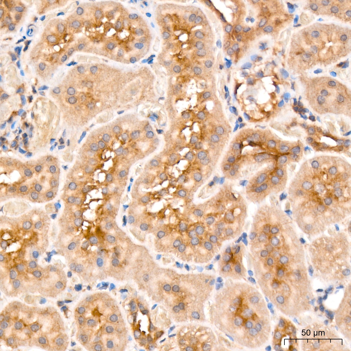
Immunohistochemistry analysis of paraffin-embedded Mouse kidney tissue using MCCC2 Rabbit pAb (CAB7990) at a dilution of 1:200 (40x lens). High pressure antigen retrieval was performed with 0.01 M citrate buffer (pH 6.0) prior to IHC staining.