Description
NAD/NADH Assay Kit (Colorimetric) (BA0067) (BA0067)
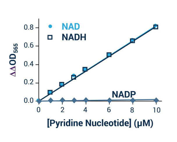
The NAD/NADH Assay Kit (Colorimetric) (SKU: BA0067) provides a simple, direct and automation-ready method for measuring NAD+ and NADH concentrations. Pyridine nucleotides play an important role in metabolism, and quantitative determination of NAD+/NADH has applications in research pertaining to energy transformation and the redox state of cells or tissue. This kit is based on a lactate dehydrogenase cycling reaction in which the formed NADH reduces a formazan (MTT) reagent; the intensity of the reduced product colour, measured at 565 nm, is proportional to the NAD+/NADH concentration. The assay is highly specific for NAD+/NADH, with minimal interference (less than 1%) from NADP+/NADPH, making it a convenient method to measure NAD, NADH and their ratio.
| Product Name: | NAD/NADH Assay Kit (Colorimetric) (BA0067) |
| SKU: | BA0067 |
| Detection Method: | Colorimetric |
| Detection Range: | 0.05 to 10 uM NAD+/NADH |
| Sample Type: | Cell or tissue extracts |
| Species Reactivity: | All |
| Assay Time: | 15 minutes |
| Kit Size: | 100 Assays |
| Equipment Required: | Microplate reader |
| Storage: | -20C |
| Shelf Life: | 6 months after receipt |
| Shipping: | Gel Pack |
Quantitative colorimetric determination of NAD+/NADH at 565 nm. A lactate dehydrogenase cycling reaction produces NADH, which reduces an MTT formazan reagent whose colour is proportional to NAD+/NADH concentration.
- Sensitive and accurate with a detection limit of 0.05 uM and linearity up to 10 uM
- Highly specific for NAD+/NADH with less than 1% interference from NADP+/NADPH
- Convenient single-working-reagent procedure with readings at time zero and 15 minutes
- High-throughput and readily automated
- Direct assays of NAD+/NADH concentrations and ratios in cell or tissue extracts
Note: The below protocol is a sample protocol. Protocols are specific to each batch/lot. For the correct instructions please follow the protocol included in your kit.
| Step | Procedure |
| 1 | Sample preparation. For tissue, weigh approximately 20 mg per sample and wash with cold PBS; for cells, wash and pellet approximately 10^5 cells. Homogenise in 100 uL NAD extraction buffer (for NAD) or 100 uL NADH extraction buffer (for NADH), heat at 60C for 5 minutes, add 20 uL Assay Buffer and 100 uL of the opposite extraction buffer to neutralise, vortex and spin at 14,000 rpm for 5 minutes; use the supernatant. |
| 2 | Calibration curve. Prepare 500 uL 10 uM NAD premix by mixing 5 uL 1 mM Standard with 495 uL distilled water and prepare the dilution series shown in the table. Transfer 40 uL standards into wells of a clear flat-bottom 96-well plate. |
| 3 | Samples. Add 40 uL of each sample into separate wells. |
| 4 | Reagent preparation. For each well, mix 60 uL Assay Buffer, 1 uL Enzyme A, 1 uL Enzyme B, 14 uL Lactate and 14 uL MTT; fresh reconstitution is recommended. |
| 5 | Reaction. Add 80 uL working reagent per well quickly and tap to mix briefly. Read optical density (OD0) at time zero at 565 nm (520-600 nm) and again (OD15) after a 15-minute incubation at room temperature. |
Compute the change in OD for each standard and sample by subtracting OD0 from OD15. Plot the standard change in OD values, determine the slope and calculate [NAD(H)] = (change in ODsample - change in ODblank) / slope x n, where n is the dilution factor. If the sample change in OD exceeds that of the 10 uM standard, dilute in distilled water, repeat and multiply by the dilution factor.
| Component | Quantity | Storage |
| Assay Buffer | 10 mL | -20C |
| Lactate | 1.5 mL | -20C |
| MTT Solution | 1.5 mL | -20C |
| Enzyme A | 120 uL | -20C |
| Enzyme B | 120 uL | -20C |
| NAD Standard | 0.5 mL | -20C |
| NAD(P)/NAD(P)H Extraction Buffers | each 12 mL | -20C |