The NDUFS8 Antibody (CAB13034) is a high-quality antibody developed for reliable detection and analysis of target proteins. This antibody, raised in rabbits, is highly specific to human samples and has been validated for Western blot applications. It binds to the NDUFV2 protein, allowing for the detection and analysis of this important mitochondrial protein in a variety of cell types.NDUFV2 is integral to the function of Complex I, which is responsible for the first step in the electron transport chain of oxidative phosphorylation. Mutations in NDUFV2 have been linked to mitochondrial diseases and disorders affecting energy production.
This antibody is validated for use in WB, IF/ICC, ELISA applications and has demonstrated reactivity against Mouse samples.
Product Name:
NDUFS8 Antibody
SKU:
CAB13034
Size:
20μL, 100μL
Reactivity:
Mouse
Conjugate:
Unconjugated
Immunogen:
Recombinant protein (or fragment).This information is considered to be commercially sensitive.
Recommended starting concentration is 1 μg/mL. Please optimize the concentration based on your specific assay requirements.
Synonyms:
TYKY, CI-23k, CI23KD, MC1DN2, NDUFS8
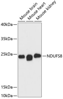
Positive Sample:
Mouse brain, Mouse heart, Mouse kidney
Cellular Localization:
Mitochondrion.
Calculated MW:
24kDa
Observed MW:
23kDa
This gene encodes a subunit of mitochondrial NADH:ubiquinone oxidoreductase, or Complex I, a multimeric enzyme of the respiratory chain responsible for NADH oxidation, ubiquinone reduction, and the ejection of protons from mitochondria. The encoded protein is involved in the binding of two of the six to eight iron-sulfur clusters of Complex I and, as such, is required in the electron transfer process. Mutations in this gene have been associated with Leigh syndrome.
Purification Method
Affinity purification
Gene ID
4728
RRID
AB_2759882
Buffer Information
Store at -20℃. Avoid freeze / thaw cycles. Buffer: PBS with 0.01% thimerosal,50% glycerol,pH7.3.
Western blot analysis of various lysates using NDUFS8 Rabbit pAb (CAB13034) at 1:3000 dilution. Secondary antibody: HRP-conjugated Goat anti-Rabbit IgG (H+L) (CABS014) at 1:10000 dilution. Lysates/proteins: 25μg per lane. Blocking buffer: 3% nonfat dry milk in TBST. Detection: ECL Basic Kit (AbGn00020). Exposure time: 90s.
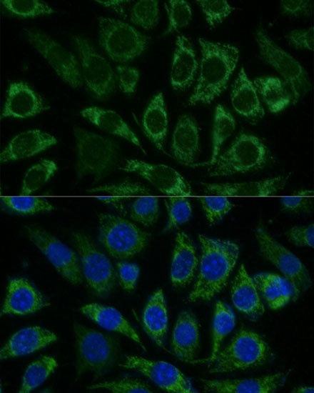
Immunofluorescence analysis of L929 cells using NDUFS8 Rabbit pAb (CAB13034) at dilution of 1:100 (40x lens). Secondary antibody: Cy3-conjugated Goat anti-Rabbit IgG (H+L) (CABS007) at 1:500 dilution. Blue: DAPI for nuclear staining.