The NRF2 Antibody (CAB0674) is a high-quality antibody developed for reliable detection and analysis of target proteins. This antibody, produced in rabbits, exhibits high reactivity with human samples and has been validated for use in Western blot applications. By binding specifically to the NFE2L2 protein, this antibody enables detection and analysis in a variety of cell types, making it ideal for investigations in oxidative stress, inflammation, and cancer research.
This antibody is validated for use in WB, IHC-P, ELISA, IF-P applications and has demonstrated reactivity against Human, Mouse, Rat samples.
Product Name:
NRF2 Antibody
SKU:
CAB0674
Size:
20μL, 100μL
Reactivity:
Human, Mouse, Rat
Conjugate:
Unconjugated
Immunogen:
Recombinant protein (or fragment).This information is considered to be commercially sensitive.
Recommended starting concentration is 1 μg/mL. Please optimize the concentration based on your specific assay requirements.
Synonyms:
NRF2, HEBP1, Nrf-2, IMDDHH
Positive Sample:
HeLa treated with MG132
Cellular Localization:
Cytoplasm, Nucleus, Cytosol.
Calculated MW:
68kDa
Observed MW:
97-110kDa
This gene encodes a transcription factor which is a member of a small family of basic leucine zipper (bZIP) proteins. The encoded transcription factor regulates genes which contain antioxidant response elements (ARE) in their promoters; many of these genes encode proteins involved in response to injury and inflammation which includes the production of free radicals. Multiple transcript variants encoding different isoforms have been characterized for this gene.
Purification Method
Affinity purification
Gene ID
4780
RRID
AB_2757326
Buffer Information
Store at -20℃. Avoid freeze / thaw cycles. Buffer: PBS with 0.09% Sodium azide,50% glycerol,pH7.3.
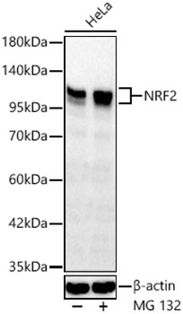
Western blot analysis of lysates from HeLa cells using NRF2 Rabbit pAb (CAB0674) at 1:1000 dilution. HeLa cells were treated with MG132(10 μM) at 37℃ for 90 minutes. Secondary antibody: HRP-conjugated Goat anti-Rabbit IgG (H+L) (CABS014) at 1:10000 dilution. Lysates/proteins: 25μg per lane. Blocking buffer: 3% nonfat dry milk in TBST. Detection: ECL Basic Kit (AbGn00020). Exposure time: 45s.
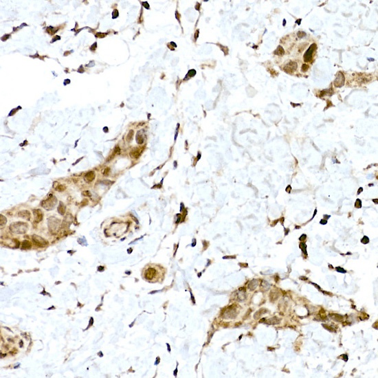
Immunohistochemistry analysis of paraffin-embedded Human breast cancer using NRF2 Rabbit pAb (CAB0674) at dilution of 1:100 (40x lens). High pressure antigen retrieval performed with 0.01M Citrate buffer (pH 6.0) prior to IHC staining.
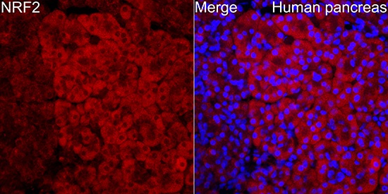
Immunofluorescence analysis of Human pancreas tissue using NRF2 Rabbit pAb (CAB0674) at a dilution of 1:200 (40x lens). Secondary antibody: Cy3-conjugated Goat anti-Rabbit IgG (H+L)(CABS007) at 1:500 dilution. Blue: DAPI for nuclear staining. High pressure antigen retrieval performed with 0.01M Citrate Buffer(pH 6.0) prior to IF staining.