The [KO Validated] PDPK1 Antibody (CAB1665) is a high-quality antibody developed for reliable detection and analysis of target proteins. This antibody, produced in rabbits, is highly specific and sensitive for detecting PDPK1 protein in human samples, making it ideal for use in Western blotting applications.PDPK1, also known as 3-phosphoinositide dependent protein kinase-1, is a crucial regulator of the PI3K/Akt signaling pathway, which is known to play a critical role in cell proliferation and survival. Dysregulation of this pathway has been implicated in various diseases, including cancer, diabetes, and cardiovascular disorders.
This antibody is validated for use in WB, IHC-P, ELISA applications and has demonstrated reactivity against Human, Mouse, Rat samples.
Product Name:
[KO Validated] PDPK1 Antibody
SKU:
CAB1665
Size:
20μL, 100μL
Reactivity:
Human, Mouse, Rat
Conjugate:
Unconjugated
Immunogen:
Recombinant protein (or fragment).This information is considered to be commercially sensitive.
Enables 3-phosphoinositide-dependent protein kinase activity; phospholipase activator activity; and phospholipase binding activity. Involved in several processes, including cell surface receptor signaling pathway; regulation of protein kinase activity; and regulation of signal transduction. Acts upstream of or within intracellular signal transduction. Located in cell projection; cytosol; and plasma membrane. Implicated in prostate cancer. Biomarker of lung non-small cell carcinoma.
Purification Method
Affinity purification
Gene ID
5170
RRID
AB_2763720
Buffer Information
Store at -20℃. Avoid freeze / thaw cycles. Buffer: PBS containing 50% glycerol, preserved with proclin300 or sodium azide, pH 7.3.
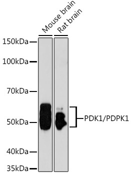
Western blot analysis of various lysates using [KO Validated] PDK1/PDPK1 Rabbit pAb (CAB1665) at 1:1000 dilution. Secondary antibody: HRP-conjugated Goat anti-Rabbit IgG (H+L) (CABS014) at 1:10000 dilution. Lysates/proteins: 25μg per lane. Blocking buffer: 3% nonfat dry milk in TBST. Detection: ECL Basic Kit (AbGn00020). Exposure time: 90s.
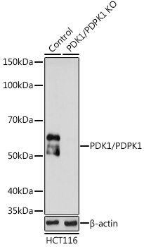
Western blot analysis of lysates from wild type (WT) and PDK1/PDPK1 knockout (KO) HCT116 cells, using [KO Validated] PDK1/PDPK1 Rabbit pAb (CAB1665) at 1:1000 dilution. Secondary antibody: HRP-conjugated Goat anti-Rabbit IgG (H+L) (CABS014) at 1:10000 dilution. Lysates/proteins: 25μg per lane. Blocking buffer: 3% nonfat dry milk in TBST. Detection: ECL Basic Kit (AbGn00020). Exposure time: 90s.