The Phospho-Histone H2AX-S139 Monoclonal Antibody (CABP0687) is a high-quality antibody developed for reliable detection and analysis of target proteins. This antibody specifically targets the phosphorylated form of histone H2AX at serine 139, a marker of double-strand DNA breaks.Phosphorylation of H2AX at serine 139 is a key event in the cellular response to DNA damage, triggering a cascade of events that recruit repair factors to the site of damage. This antibody, raised in rabbits, is highly sensitive and specific for detecting phosphorylated H2AX in various cell types and is validated for use in Western blot and immunofluorescence applications.
This antibody is validated for use in WB, IHC-P, IF/ICC, ELISA applications and has demonstrated reactivity against Human, Mouse, Rat samples.
Product Name:
Phospho-Histone H2AX-S139 Monoclonal Antibody
SKU:
CABP0687
Size:
20μL, 100μL
Reactivity:
Human, Mouse, Rat
Clone Number:
ARC0110
Conjugate:
Unconjugated
Immunogen:
Synthetic peptide. This information is considered to be commercially sensitive.
Sequence:
TQAS Q
Tested Applications:
WBIHC-PIF/ICCELISA
Recommended Dilution:
WB
1:7000 - 1:39000
IHC-P
1:500 - 1:2000
IF/ICC
1:1000 - 1:5000
ELISA
Recommended starting concentration is 1 μg/mL. Please optimize the concentration based on your specific assay requirements.
Synonyms:
H2A.X, H2A/X, H2AFX, Phospho-Histone H2AX-S139
Positive Sample:
293T treated with UV, NIH/3T3 treated with UV, C6 treated with UV
Cellular Localization:
Chromosome, Nucleus.
Calculated MW:
15kDa
Observed MW:
15kDa
Histones are basic nuclear proteins that are responsible for the nucleosome structure of the chromosomal fiber in eukaryotes. Two molecules of each of the four core histones (H2A, H2B, H3, and H4) form an octamer, around which approximately 146 bp of DNA is wrapped in repeating units, called nucleosomes. The linker histone, H1, interacts with linker DNA between nucleosomes and functions in the compaction of chromatin into higher order structures. This gene encodes a replication-independent histone that is a member of the histone H2A family, and generates two transcripts through the use of the conserved stem-loop termination motif, and the polyA addition motif.
Purification Method
Affinity purification
Gene ID
3014
RRID
AB_2863808
Buffer Information
Store at -20℃. Avoid freeze / thaw cycles. Buffer: PBS with 0.09% Sodium azide,0.05% BSA,50% glycerol,pH7.3.
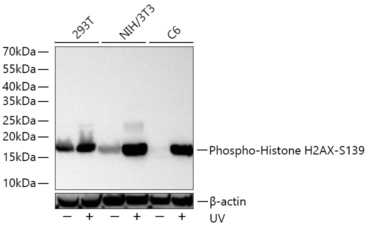
Western blot analysis of various lysates using Phospho-Histone H2AX-S139 Rabbit mAb (CABP0687) at 1:7000 dilution incubated overnight at 4℃. 293T,NIH/3T3 and C6 cells were treated with UV at room temperature for 15-30 minutes. Secondary antibody: HRP-conjugated Goat anti-Rabbit IgG (H+L) (CABS014) at 1:10000 dilution. Lysates/proteins: 30 μg per lane. Blocking buffer: 3% nonfat dry milk in TBST. Detection: ECL Basic Kit (AbGn00020). Exposure time: 20 s.
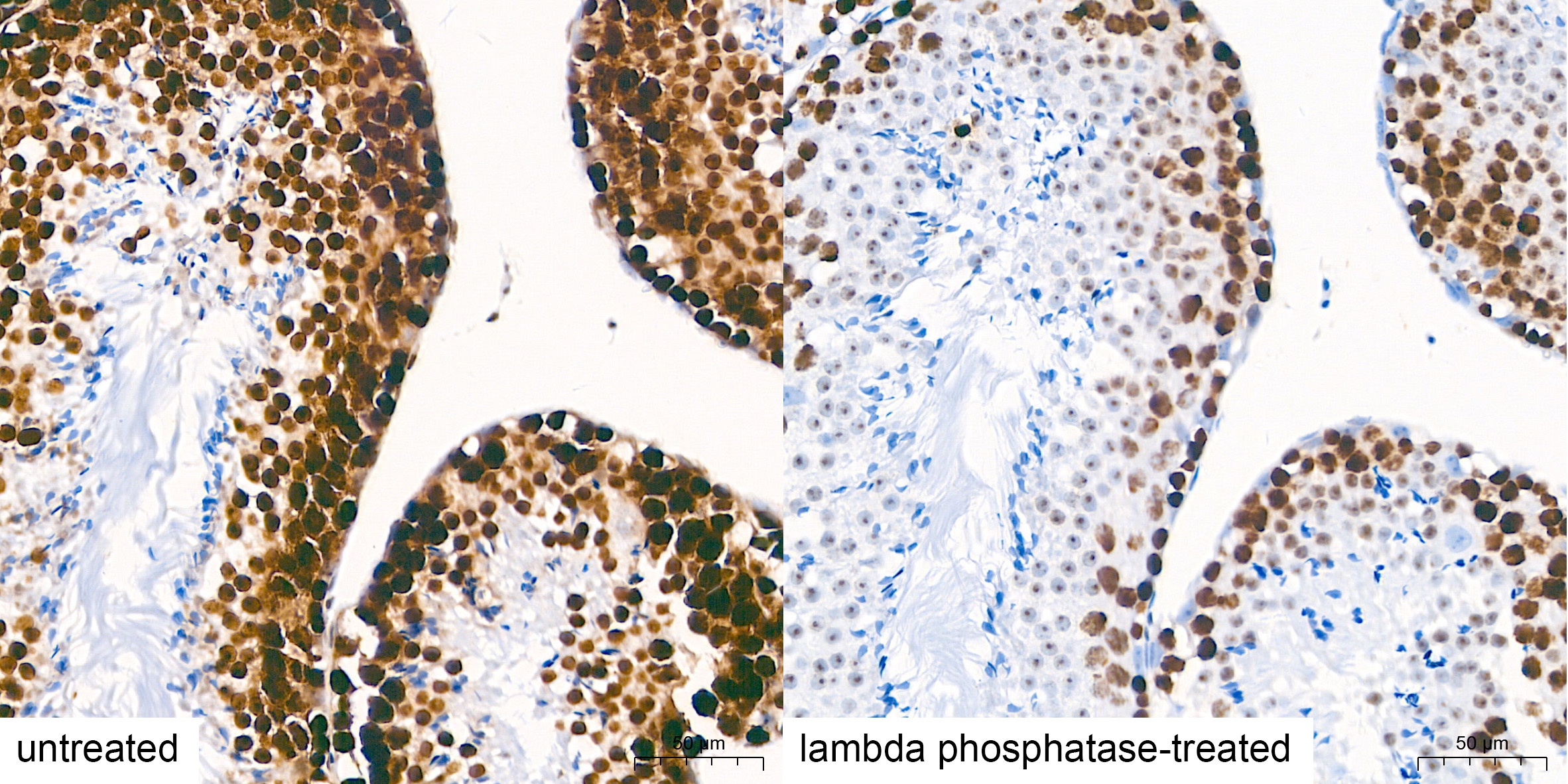
Immunohistochemistry analysis of paraffin-embedded Mouse testis tissue using Phospho-Histone H2AX-S139 Rabbit mAb (CABP0687) at a dilution of 1:1000 (40x lens). High pressure antigen retrieval performed with 0.01M Citrate Buffer (pH 6.0) prior to IHC staining.
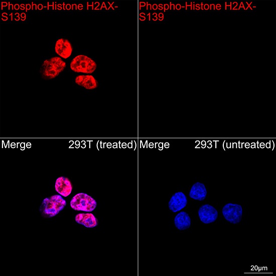
Confocal imaging of 293T cells (treated with UV) and 293T cells (untreated) using Phospho-Histone H2AX-S139 Rabbit mAb (CABP0687, dilution 1:5000) followed by a further incubation with Cy3 Goat Anti-Rabbit IgG (H+L) (CABS007, dilution 1:500) (Red). DAPI was used for nuclear staining (Blue). Objective: 100x.
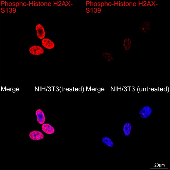
Confocal imaging of NIH/3T3 cells (treated with UV) and NIH/3T3 cells (untreated) using Phospho-Histone H2AX-S139 Rabbit mAb (CABP0687, dilution 1:5000) followed by a further incubation with Cy3 Goat Anti-Rabbit IgG (H+L) (CABS007, dilution 1:500) (Red). DAPI was used for nuclear staining (Blue). Objective: 100x.