The Phospho-Histone H3-S28 Antibody (CABP0839) is a high-quality antibody developed for reliable detection and analysis of target proteins. This antibody, produced in rabbits, is highly specific for detecting phosphorylation of Histone H3 at Serine 28 in human samples. Validated for use in Western blotting applications, this antibody allows for the precise detection and analysis of phosphorylated Histone H3 in various cell types.Histone H3 is a core histone protein that plays a crucial role in chromatin structure and gene regulation. Phosphorylation of Histone H3 at Serine 28 is known to be involved in mitotic chromosome condensation and cell cycle progression, making it a key marker for studying cell division and proliferation processes.
This antibody is validated for use in WB, IF/ICC, ELISA applications and has demonstrated reactivity against Human, Mouse, Rat, Other (Wide Range Predicted) samples.
Product Name:
Phospho-Histone H3-S28 Antibody
SKU:
CABP0839
Size:
20μL, 100μL
Reactivity:
Human, Mouse, Rat, Other (Wide Range Predicted)
Conjugate:
Unconjugated
Immunogen:
Synthetic peptide. This information is considered to be commercially sensitive.
Sequence:
ARKS A
Tested Applications:
WBIF/ICCELISA
Recommended Dilution:
WB
1:500 - 1:2000
IF/ICC
1:50 - 1:200
ELISA
Recommended starting concentration is 1 μg/mL. Please optimize the concentration based on your specific assay requirements.
HeLa treated with Calyculin A, NIH/3T3 treated with Calyculin A, C6 treated with Calyculin A
Cellular Localization:
Chromosome, Nucleus.
Calculated MW:
15kDa
Observed MW:
17kDa
Histones are basic nuclear proteins that are responsible for the nucleosome structure of the chromosomal fiber in eukaryotes. This structure consists of approximately 146 bp of DNA wrapped around a nucleosome, an octamer composed of pairs of each of the four core histones (H2A, H2B, H3, and H4). The chromatin fiber is further compacted through the interaction of a linker histone, H1, with the DNA between the nucleosomes to form higher order chromatin structures. This gene is intronless and encodes a replication-dependent histone that is a member of the histone H3 family. Transcripts from this gene lack polyA tails; instead, they contain a palindromic termination element. This gene is found in the large histone gene cluster on chromosome 6p22-p21.3.
Purification Method
Affinity purification
Gene ID
8290 8350
RRID
AB_2771173
Buffer Information
Store at -20℃. Avoid freeze / thaw cycles. Buffer: PBS containing 50% glycerol, preserved with proclin300 or sodium azide, pH 7.3.
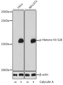
Western blot analysis of various lysates using Phospho-Histone H3-S28 Rabbit pAb (CABP0839) at 1:1000 dilution. Both HeLa cells and NIH/3T3 cells were treated with Calyculin A (100 nM) at 37℃ for 30 minutes after serum-starvation overnight. Secondary antibody: HRP-conjugated Goat anti-Rabbit IgG (H+L) (CABS014) at 1:10000 dilution. Lysates/proteins: 25μg per lane. Blocking buffer: 3% BSA. Detection: ECL Basic Kit (AbGn00020). Exposure time: 1s.
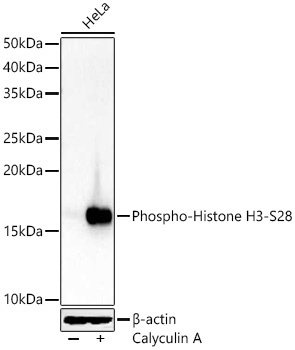
Western blot analysis of lysates from HeLa cells, using Phospho-Histone H3-S28 Rabbit pAb (CABP0839) at 1:900 dilution. HeLa cells were treated with Calyculin A (100 nM) at 37℃ for 30 minutes after serum-starvation overnight. Secondary antibody: HRP-conjugated Goat anti-Rabbit IgG (H+L) (CABS014) at 1:10000 dilution. Lysates/proteins: 25μg per lane. Blocking buffer: 3% nonfat dry milk in TBST. Detection: ECL Basic Kit (AbGn00020). Exposure time: 10s.
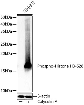
Western blot analysis of lysates from NIH/3T3 cells, using Phospho-Histone H3-S28 Rabbit pAb (CABP0839) at 1:900 dilution. NIH/3T3 cells were treated with Calyculin A (100 nM) at 37℃ for 30 minutes after serum-starvation overnight. Secondary antibody: HRP-conjugated Goat anti-Rabbit IgG (H+L) (CABS014) at 1:10000 dilution. Lysates/proteins: 25μg per lane. Blocking buffer: 3% nonfat dry milk in TBST. Detection: ECL Basic Kit (AbGn00020). Exposure time: 0.5s.
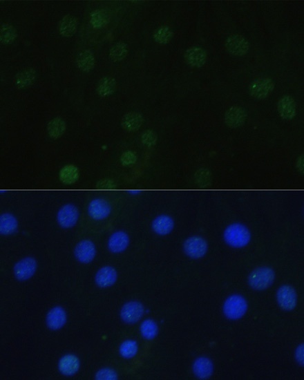
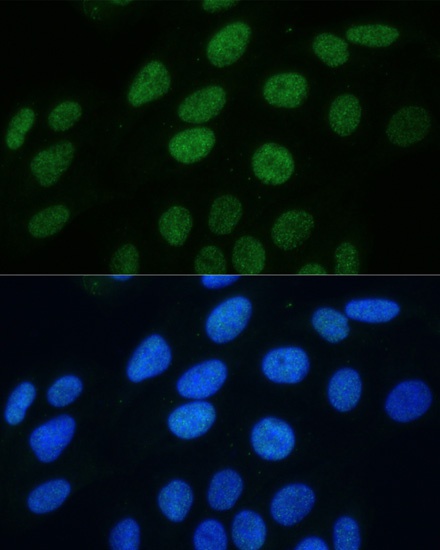
Immunofluorescence analysis of H9C2 cells using Phospho-Histone H3-S28 Rabbit pAb (CABP0839) at dilution of 1:100. Blue: DAPI for nuclear staining.
Immunofluorescence analysis of U2OS cells using Phospho-Histone H3-S28 Rabbit pAb (CABP0839) at dilution of 1:100. Blue: DAPI for nuclear staining.