The Phospho-MAPKAPK2-T334 Antibody (CABP0588) is a high-quality antibody developed for reliable detection and analysis of target proteins. MAPKAPK2 is a protein kinase involved in the MAPK signaling pathway, which plays a key role in cell proliferation, differentiation, and apoptosis. Phosphorylation of MAPKAPK2 at T334 is known to regulate its activity and substrate specificity.This antibody, produced in rabbits, shows high reactivity with human samples and has been validated for use in Western blot applications. It specifically binds to the phosphorylated form of MAPKAPK2 at T334, enabling researchers to detect and analyze the phosphorylation status of this protein in various cell types.
This antibody is validated for use in WB, ELISA applications and has demonstrated reactivity against Human, Mouse, Rat samples.
Product Name:
Phospho-MAPKAPK2-T334 Antibody
SKU:
CABP0588
Size:
20μL, 100μL
Reactivity:
Human, Mouse, Rat
Conjugate:
Unconjugated
Immunogen:
Synthetic peptide. This information is considered to be commercially sensitive.
Sequence:
PQTP L
Tested Applications:
WBELISA
Recommended Dilution:
WB
1:500 - 1:1000
ELISA
Recommended starting concentration is 1 μg/mL. Please optimize the concentration based on your specific assay requirements.
Synonyms:
MK2, MK-2, MAPKAP-K2, Phospho-MAPKAPK-2/MK2-T334
Positive Sample:
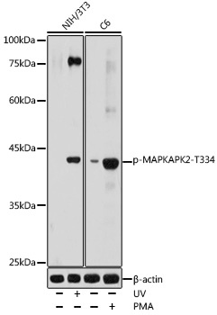
NIH/3T3 treated with UV, C6 treated with PMA
Cellular Localization:
Cytoplasm, Nucleus.
Calculated MW:
46kDa
Observed MW:
42kDa
This gene encodes a member of the Ser/Thr protein kinase family. This kinase is regulated through direct phosphorylation by p38 MAP kinase. In conjunction with p38 MAP kinase, this kinase is known to be involved in many cellular processes including stress and inflammatory responses, nuclear export, gene expression regulation and cell proliferation. Heat shock protein HSP27 was shown to be one of the substrates of this kinase in vivo. Two transcript variants encoding two different isoforms have been found for this gene.
Purification Method
Affinity purification
Gene ID
9261
RRID
AB_2771319
Buffer Information
Store at -20℃. Avoid freeze / thaw cycles. Buffer: PBS with 0.09% Sodium azide,50% glycerol,pH7.3.
Western blot analysis of various lysates using Phospho-MAPKAPK-2/MK2-T334 Rabbit pAb (CABP0588) at 1:1000 dilution. NIH/3T3 cells were treated with UV at room temperature for 15-30 minutes. C6 cells were treated with PMA/TPA (200 nM) at 37℃ for 30 minutes after serum-starvation overnight. Secondary antibody: HRP-conjugated Goat anti-Rabbit IgG (H+L) (CABS014) at 1:10000 dilution. Lysates/proteins: 25μg per lane. Blocking buffer: 3% nonfat dry milk in TBST. Detection: ECL Basic Kit (AbGn00020). Exposure time: 180s.