The [KO Validated] SCD Antibody (CAB16429) is a high-quality antibody developed for reliable detection and analysis of target proteins. This antibody, derived from rabbits, demonstrates high reactivity with human samples and is suitable for Western blot applications. By binding to SCD, this antibody allows for the detection and analysis of the enzyme in various cell types, making it ideal for investigations in the fields of metabolism and lipid metabolism research.SCD plays a vital role in lipid metabolism by catalyzing the desaturation of saturated fatty acids to monounsaturated fatty acids, which are not only essential for membrane structure but also play key roles in lipid signaling pathways.
This antibody is validated for use in WB, IHC-P, IF/ICC, ELISA applications and has demonstrated reactivity against Human, Mouse, Rat samples.
Product Name:
[KO Validated] SCD Antibody
SKU:
CAB16429
Size:
20μL, 100μL
Reactivity:
Human, Mouse, Rat
Conjugate:
Unconjugated
Immunogen:
Synthetic peptide. This information is considered to be commercially sensitive.
This gene encodes an enzyme involved in fatty acid biosynthesis, primarily the synthesis of oleic acid. The protein belongs to the fatty acid desaturase family and is an integral membrane protein located in the endoplasmic reticulum. Transcripts of approximately 3.9 and 5.2 kb, differing only by alternative polyadenlyation signals, have been detected. A gene encoding a similar enzyme is located on chromosome 4 and a pseudogene of this gene is located on chromosome 17.
Purification Method
Affinity purification
Gene ID
6319
RRID
AB_2772150
Buffer Information
Store at -20℃. Avoid freeze / thaw cycles. Buffer: PBS with 0.09% Sodium azide,50% glycerol,pH7.3.
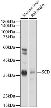
Western blot analysis of various lysates using SCD Rabbit pAb (CAB16429) at 1:1000 dilution. Secondary antibody: HRP-conjugated Goat anti-Rabbit IgG (H+L) (CABS014) at 1:10000 dilution. Lysates/proteins: 25μg per lane. Blocking buffer: 3% nonfat dry milk in TBST. Detection: ECL Basic Kit (AbGn00020). Exposure time: 1s.
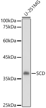
Western blot analysis of lysates from U-251MG cells, using SCD Rabbit pAb (CAB16429) at 1:1000 dilution. Secondary antibody: HRP-conjugated Goat anti-Rabbit IgG (H+L) (CABS014) at 1:10000 dilution. Lysates/proteins: 25μg per lane. Blocking buffer: 3% nonfat dry milk in TBST. Detection: ECL Basic Kit (AbGn00020). Exposure time: 3s.
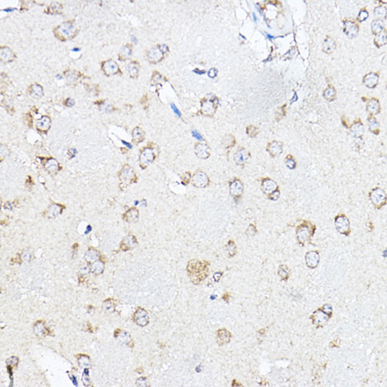
Immunohistochemistry analysis of paraffin-embedded Mouse brain using SCD Rabbit pAb (CAB16429) at dilution of 1:100 (40x lens). High pressure antigen retrieval performed with 0.01M Citrate buffer (pH 6.0) prior to IHC staining.
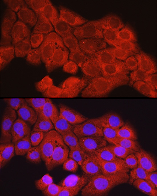
Immunofluorescence analysis of A431 cells using SCD Rabbit pAb (CAB16429) at dilution of 1:100 (40x lens). Secondary antibody: Cy3-conjugated Goat anti-Rabbit IgG (H+L) (CABS007) at 1:500 dilution. Blue: DAPI for nuclear staining.