The SOCS3 Monoclonal Antibody (CAB21981) is a high-quality antibody developed for reliable detection and analysis of target proteins. This monoclonal antibody, produced through advanced technology, is highly specific and sensitive for detecting SOCS3 in various biological samples. SOCS3 is known for its ability to inhibit the JAK/STAT pathway, a key signaling pathway involved in immune response and inflammation. Dysregulation of SOCS3 has been linked to various diseases such as cancer, autoimmune disorders, and inflammatory conditions. By studying the expression and function of SOCS3, researchers can gain valuable insights into the mechanisms underlying these diseases and potentially identify new therapeutic targets.
This antibody is validated for use in WB, ELISA applications and has demonstrated reactivity against Mouse samples.
Product Name:
SOCS3 Monoclonal Antibody
SKU:
CAB21981
Size:
20μL, 100μL
Reactivity:
Mouse
Clone Number:
ARC53312
Conjugate:
Unconjugated
Immunogen:
Synthetic peptide. This information is considered to be commercially sensitive.
Recommended starting concentration is 1 μg/mL. Please optimize the concentration based on your specific assay requirements.
Synonyms:
CIS3, SSI3, ATOD4, Cish3, SSI-3, SOCS-3, SOCS3
Positive Sample:
RAW 264.7 treated with LPS
Cellular Localization:
Cytosol.
Calculated MW:
25kDa
Observed MW:
28kDa
This gene encodes a member of the STAT-induced STAT inhibitor (SSI), also known as suppressor of cytokine signaling (SOCS), family. SSI family members are cytokine-inducible negative regulators of cytokine signaling. The expression of this gene is induced by various cytokines, including IL6, IL10, and interferon (IFN)-gamma. The protein encoded by this gene can bind to JAK2 kinase, and inhibit the activity of JAK2 kinase. Studies of the mouse counterpart of this gene suggested the roles of this gene in the negative regulation of fetal liver hematopoiesis, and placental development.
Purification Method
Affinity purification
Gene ID
9021
Buffer Information
Store at -20℃. Avoid freeze / thaw cycles. Buffer: PBS containing 50% glycerol and 0.05% BSA, preserved with proclin300 or sodium azide, pH 7.3.
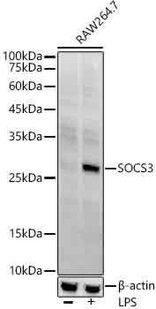
Western blot analysis of lysates from RAW 264.7 cells using SOCS3 Rabbit mAb (CAB21981) at1:2000 dilution incubated overnight at 4℃. Raw 264. 7 cells were treated with LPS (1 μg/ml) at 37℃ for 8 hours. Secondary antibody: HRP-conjugated Goat anti-Rabbit IgG (H+L) (CABS014) at 1:10000 dilution. Lysates/proteins: 25 μg per lane. Blocking buffer: 3% nonfat dry milk in TBST. Detection: ECL Enhanced Kit (AbGn00021). Exposure time: 180 s.
 at1:2000 dilution. Raw264. 7 cells were treated by LPS (1 μg/ml) at 37℃ for 8 hours. Secondary antibody: HRP Goat Anti-Rabbit IgG (H+L) at 1:10000 dilution. Lysates/proteins: 25μg per lane. Blocking buffer: 3% nonfat dry milk in TBST.")
 at1:2000 dilution. Raw264. 7 cells were treated by LPS (1 μg/ml) at 37℃ for 8 hours. Secondary antibody: HRP Goat Anti-Rabbit IgG (H+L) at 1:10000 dilution. Lysates/proteins: 25μg per lane. Blocking buffer: 3% nonfat dry milk in TBST.")

 at 1:10000 dilution. Lysates/proteins: 25ug per lane. Blocking buffer: 3% nonfat dry milk in TBST. Detection: ECL Basic Kit. Exposure time: 180s.")
 at 1:10000 dilution. Lysates/proteins: 25ug per lane. Blocking buffer: 3% nonfat dry milk in TBST. Detection: ECL Basic Kit. Exposure time: 90s.")
 at 1:10000 dilution. Lysates/proteins: 25ug per lane. Blocking buffer: 3% nonfat dry milk in TBST. Detection: ECL Basic Kit. Exposure time: 180s.")
 at 1:10000 dilution. Lysates/proteins: 25ug per lane. Blocking buffer: 3% nonfat dry milk in TBST. Detection: ECL Basic Kit. Exposure time: 30s.")
 at 1:10000 dilution. Lysates/proteins: 25ug per lane. Blocking buffer: 3% nonfat dry milk in TBST. Detection: ECL Basic Kit. Exposure time: 60s.")
. Perform high pressure antigen retrieval with 10 mM citrate buffer pH 6. 0 before commencing with IHC staining protocol.")
. Perform high pressure antigen retrieval with 10 mM citrate buffer pH 6. 0 before commencing with IHC staining protocol.")