The Thioredoxin 1 (Trx1/TXN) Monoclonal Antibody (CAB4024) is a high-quality antibody developed for reliable detection and analysis of target proteins. This antibody, specifically raised in rabbits, is highly reactive with human samples and has been validated for use in a variety of applications, including Western blotting and immunofluorescence.TRX1, also known as thioredoxin 1, plays a vital role in the regulation of cellular redox balance and protection against oxidative stress. Its importance in maintaining cellular health makes it a valuable target for research in fields such as cancer, neurobiology, and cardiovascular disease.
This antibody is validated for use in WB, IHC-P, ELISA applications and has demonstrated reactivity against Human, Mouse, Rat samples.
Product Name:
Thioredoxin 1 (Trx1/TXN) Monoclonal Antibody
SKU:
CAB4024
Size:
20μL, 100μL
Reactivity:
Human, Mouse, Rat
Clone Number:
ARC2658
Conjugate:
Unconjugated
Immunogen:
Synthetic peptide. This information is considered to be commercially sensitive.
Recommended starting concentration is 1 μg/mL. Please optimize the concentration based on your specific assay requirements.
Synonyms:
TRX, TRDX, TRX1, Trx80, Thioredoxin 1 (Trx1/TXN)
Positive Sample:
HeLa, MCF7, C2C12, Mouse lung, Rat brain
Cellular Localization:
Cytoplasm, Nucleus, Secreted.
Calculated MW:
12kDa
Observed MW:
12kDa
The protein encoded by this gene acts as a homodimer and is involved in many redox reactions. The encoded protein is active in the reversible S-nitrosylation of cysteines in certain proteins, which is part of the response to intracellular nitric oxide. This protein is found in the cytoplasm. Two transcript variants encoding different isoforms have been found for this gene.
Purification Method
Affinity purification
Gene ID
7295
RRID
AB_2863174
Buffer Information
Store at -20℃. Avoid freeze / thaw cycles. Buffer: PBS containing 50% glycerol and 0.05% BSA, preserved with proclin300 or sodium azide, pH 7.3.
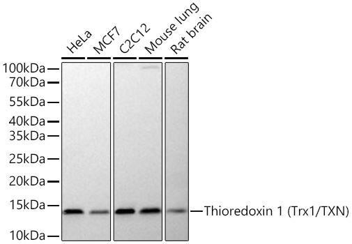
Western blot analysis of various lysates using Thioredoxin 1 (Trx1/TXN) Rabbit mAb (CAB4024) at 1:10000 dilution incubated at room temperature for 1.5 hours. Secondary antibody: HRP-conjugated Goat anti-Rabbit IgG (H+L) (CABS014) at 1:10000 dilution. Lysates/proteins: 25 μg per lane. Blocking buffer: 3% nonfat dry milk in TBST. Detection: ECL Basic Kit (AbGn00020). Exposure time: 90 s.
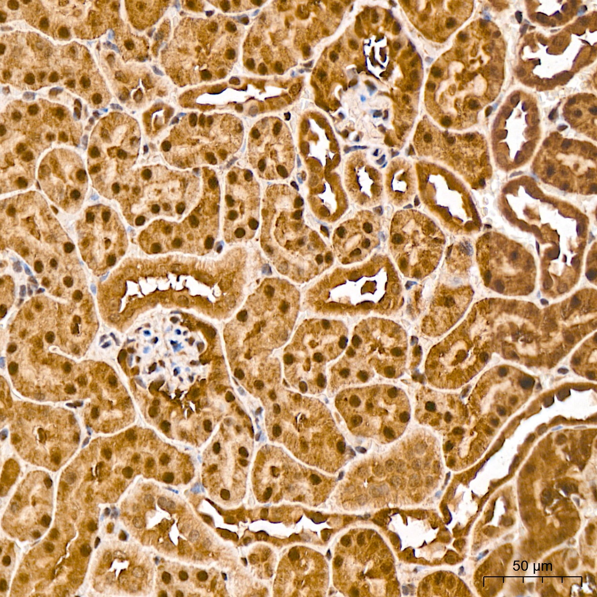
Immunohistochemistry analysis of paraffin-embedded Mouse kidney tissue using Thioredoxin 1 (Trx1/TXN) Rabbit mAb (CAB4024) at a dilution of 1:1600 (40x lens). High pressure antigen retrieval was performed with 0.01 M citrate buffer (pH 6.0) prior to IHC staining.
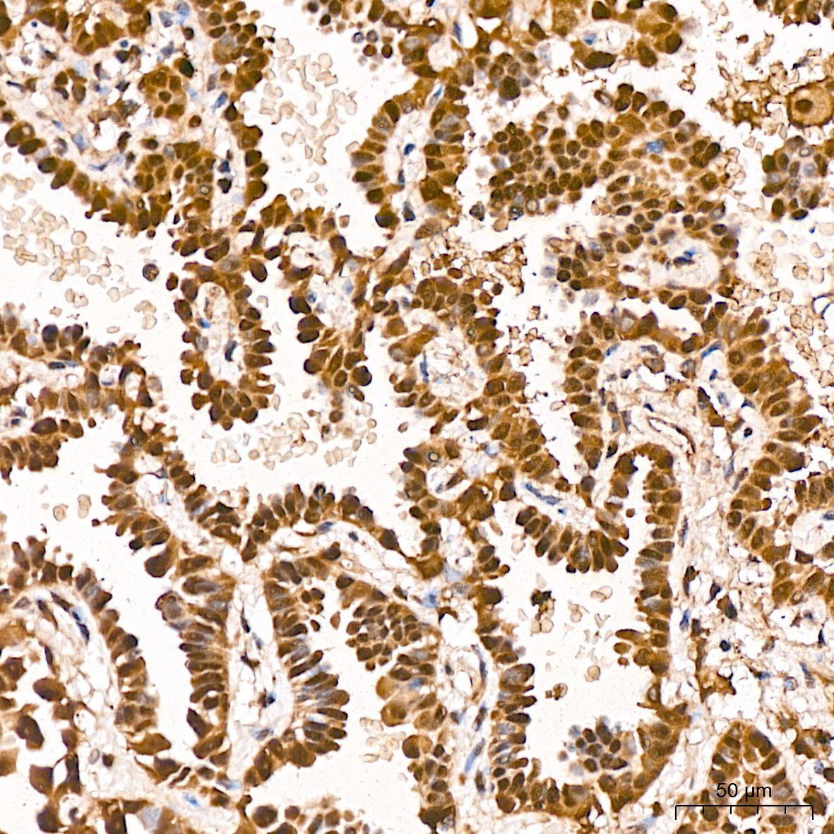
Immunohistochemistry analysis of paraffin-embedded Human lung adenocarcinoma tissue using Thioredoxin 1 (Trx1/TXN) Rabbit mAb (CAB4024) at a dilution of 1:1600 (40x lens). High pressure antigen retrieval was performed with 0.01 M citrate buffer (pH 6.0) prior to IHC staining.


 ELISA Kit (AEFI00818)")
 ELISA Kit (AEFI00818)")


