


Description
ACHE Antibody (CAB2806)
The ACHE Antibody (CAB2806) is a high-quality antibody developed for reliable detection and analysis of target proteins. This antibody, produced in rabbits, is highly specific to human samples and is validated for use in Western blot applications.AChE is primarily found in the nervous system and at neuromuscular junctions, where it controls nerve signal transmission. Dysregulation of AChE activity has been linked to various neurological disorders, including Alzheimer's disease and myasthenia gravis. Therefore, studying AChE expression and function is essential for understanding these conditions and developing potential treatments.
This antibody is validated for use in WB, IF/ICC, ELISA applications and has demonstrated reactivity against Human, Mouse, Rat samples.
| Product Name: | ACHE Antibody |
| SKU: | CAB2806 |
| Size: | 20μL, 100μL |
| Reactivity: | Human, Mouse, Rat |
| Conjugate: | Unconjugated |
| Immunogen: | Recombinant protein (or fragment).This information is considered to be commercially sensitive. | ||||||
| Sequence: | VPQV SDLA AEAV VLHY TDWL HPED PARL REAL SDVV GDHN VVCP VAQL AGRL AAQG ARVY AYVF EHRA STLS WPLW MGVP HGYE IEFI FGIP LDPS RNYT AEEK IFAQ RLMR YWAN FART GDPN EPRD PKAP QWPP YTAG AQQY VSLD LRPL EVRR GLRA QACA FWNR FLPK LLSA TASE APST CPGF THGE AAPR PGLP LPLL LLHQ LLLL FLSH LRRL | ||||||
| Tested Applications: | WB IF/ICC ELISA | ||||||
| Recommended Dilution: |
| ||||||
| Synonyms: | YT, ACEE, ARACHE, N-ACHE, ACHE |
| Positive Sample: | SH-SY5Y, SGC996, Mouse brain, Mouse spinal cord, Mouse liver, Rat spinal cord |
| Cellular Localization: | Cell Junction, Cell Membrane, Extracellular Side, Gpi-Anchor, Lipid-Anchor, Nucleus, Peripheral Membrane Protein, Secreted, Synapse. |
| Calculated MW: | 68kDa |
| Observed MW: | 75kDa |
Acetylcholinesterase hydrolyzes the neurotransmitter, acetylcholine at neuromuscular junctions and brain cholinergic synapses, and thus terminates signal transmission. It is also found on the red blood cell membranes, where it constitutes the Yt blood group antigen. Acetylcholinesterase exists in multiple molecular forms which possess similar catalytic properties, but differ in their oligomeric assembly and mode of cell attachment to the cell surface. It is encoded by the single ACHE gene, and the structural diversity in the gene products arises from alternative mRNA splicing, and post-translational associations of catalytic and structural subunits. The major form of acetylcholinesterase found in brain, muscle and other tissues is the hydrophilic species, which forms disulfide-linked oligomers with collagenous, or lipid-containing structural subunits. The other, alternatively spliced form, expressed primarily in the erythroid tissues, differs at the C-terminal end, and contains a cleavable hydrophobic peptide with a GPI-anchor site. It associates with the membranes through the phosphoinositide (PI) moieties added post-translationally. AChE activity may constitute a sensitive biomarker of RBC ageing in vivo, and thus, may be of aid in understanding the effects of transfusion
| Purification Method | Affinity purification |
| Gene ID | 43 |
| RRID | AB_2764644 |
| Buffer Information | Store at -20℃. Avoid freeze / thaw cycles. Buffer: PBS containing 50% glycerol, preserved with proclin300 or sodium azide, pH 7.3. |
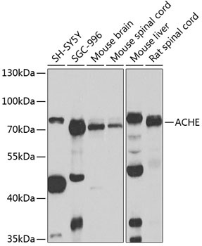 | Western blot analysis of various lysates using ACHE Rabbit pAb (CAB2806) at 1:1000 dilution. Secondary antibody: HRP-conjugated Goat anti-Rabbit IgG (H+L) (CABS014) at 1:10000 dilution. Lysates/proteins: 25μg per lane. Blocking buffer: 3% nonfat dry milk in TBST. Detection: ECL Basic Kit (AbGn00020). Exposure time: 90s. |
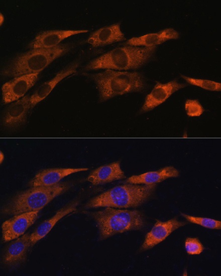 | Immunofluorescence analysis of NIH/3T3 cells using ACHE Rabbit pAb (CAB2806) at dilution of 1:100. Secondary antibody: Cy3-conjugated Goat anti-Rabbit IgG (H+L) (CABS007) at 1:500 dilution. Blue: DAPI for nuclear staining. |
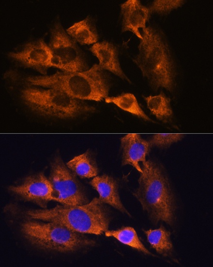 | Immunofluorescence analysis of C6 cells using ACHE Rabbit pAb (CAB2806) at dilution of 1:100. Secondary antibody: Cy3-conjugated Goat anti-Rabbit IgG (H+L) (CABS007) at 1:500 dilution. Blue: DAPI for nuclear staining. |


