Description
ADP Assay Kit (Fluorometric) (BA0063) (BA0063)
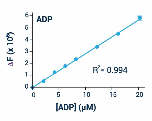
The ADP Assay Kit (Fluorometric) (SKU: BA0063) provides a convenient fluorometric means of measuring adenosine diphosphate (ADP) levels, even in the presence of ATP. ADP is the product of ATP dephosphorylation by ATPases and can be converted back to ATP by ATP synthases, with ADP levels regulating several enzymes involved in intermediary metabolism. Conventional luciferase/luciferin assays measure ADP after conversion to ATP, but the nascent ATP signal can drown out the ADP signal; this newly designed assay overcomes that limitation. In the assay, ADP is converted to ATP and pyruvate, and the generated pyruvate is quantified fluorometrically at excitation/emission 530/590 nm. The assay is simple, sensitive, stable and high-throughput adaptable, detecting as little as 0.1 uM ADP in biological samples.
| Product Name: | ADP Assay Kit (Fluorometric) (BA0063) |
| SKU: | BA0063 |
| Detection Method: | Fluorometric |
| Detection Range: | As low as 0.1 uM; up to 20 uM ADP |
| Sample Type: | Cells and other biological samples |
| Species Reactivity: | All |
| Assay Time: | 30 minutes |
| Kit Size: | 100 Assays |
| Equipment Required: | Microplate reader |
| Storage: | -20C |
| Shelf Life: | At least 6 months |
| Shipping: | Gel Pack |
Quantitative fluorometric determination of adenosine diphosphate. ADP is converted to ATP and pyruvate, and the generated pyruvate is quantified fluorometrically at excitation/emission 530/590 nm.
- Safe, non-radioactive assay
- Sensitive: as low as 0.1 uM ADP can be quantified
- Homogeneous, convenient mix-incubate-measure format with no wash or transfer steps
- Robust and amenable to high-throughput screening
- ADP determination in cells and other biological samples
Note: The below protocol is a sample protocol. Protocols are specific to each batch/lot. For the correct instructions please follow the protocol included in your kit.
| Step | Procedure |
| 1 | Standards. Use black flat-bottom plates and bring all reagents to room temperature. Prepare 900 uL 20 uM ADP premix by mixing 6 uL 3 mM Standard with 894 uL distilled water and prepare the dilution series shown in the table. Transfer 40 uL standards into separate wells. |
| 2 | Sample preparation. Samples high in protein or with likely ATPase activity should be deproteinated and neutralised: add 25 uL 10% TCA per 100 uL sample, vortex, centrifuge 10 minutes at 14,000 rpm, transfer 100 uL clear supernatant and neutralise with 12.5 uL Neutralizer. Multiply the measured change in RFU for deproteinated samples by 1.41 to compensate for dilution. |
| 3 | Transfer 40 uL of each sample into separate wells; for samples containing pyruvate, add to two wells so one can serve as the sample blank. |
| 4 | Prepare working reagent for each well by mixing 45 uL Reagent A, 45 uL Reagent B and 1 uL Enzyme (omit enzyme for sample blanks). Add 80 uL of the appropriate working reagent to each well, tap to mix and incubate at room temperature for 30 minutes protected from light. |
| 5 | Read fluorescence intensity at excitation 530 nm and emission 590 nm. |
Plot the RFU measured at 30 minutes for each standard against the standard concentrations and determine the slope by linear regression. [ADP] = (RFUsample - RFUblank) / slope x n, where n is the sample dilution factor (1.41 for deproteinated samples). If the ADP concentration exceeds 20 uM, dilute the sample in water, repeat and multiply by the dilution factor.
| Component | Quantity | Storage |
| Reagent A | 6 mL | -20C |
| Reagent B | 6 mL | -20C |
| Enzyme | 120 uL | -20C |
| Standard | 100 uL | -20C |
| 10% TCA | 6 mL | -20C |
| Neutralizer | 1.5 mL | -20C |