The CD38 Antibody (CAB13611) is a high-quality antibody developed for reliable detection and analysis of target proteins. Produced using rabbit immunization methods, this antibody has high reactivity with human samples and has been extensively validated for Western blot applications.CD38, a multifunctional enzyme involved in cell signaling and immune regulation, is a promising target for research in immunology, oncology, and autoimmune diseases. By detecting and analyzing CD38 protein levels in various cell types, this antibody enables researchers to gain insights into its functions and potential therapeutic implications.
This antibody is validated for use in WB, IHC-P, IF/ICC, ELISA applications and has demonstrated reactivity against Human, Mouse, Rat samples.
Product Name:
CD38 Antibody
SKU:
CAB13611
Size:
20μL, 100μL
Reactivity:
Human, Mouse, Rat
Conjugate:
Unconjugated
Immunogen:
Recombinant protein (or fragment).This information is considered to be commercially sensitive.
Recommended starting concentration is 1 μg/mL. Please optimize the concentration based on your specific assay requirements.
Synonyms:
ADPRC1, cADPR1, ADPRC 1, CD38
Positive Sample:
Ramos
Cellular Localization:
Membrane, Single-Pass Type Ii Membrane Protein.
Calculated MW:
34kDa
Observed MW:
45kDa
The protein encoded by this gene is a non-lineage-restricted, type II transmembrane glycoprotein that synthesizes and hydrolyzes cyclic adenosine 5'-diphosphate-ribose, an intracellular calcium ion mobilizing messenger. The release of soluble protein and the ability of membrane-bound protein to become internalized indicate both extracellular and intracellular functions for the protein. This protein has an N-terminal cytoplasmic tail, a single membrane-spanning domain, and a C-terminal extracellular region with four N-glycosylation sites. Crystal structure analysis demonstrates that the functional molecule is a dimer, with the central portion containing the catalytic site. It is used as a prognostic marker for patients with chronic lymphocytic leukemia. Alternative splicing results in multiple transcript variants.
Purification Method
Affinity purification
Gene ID
952
RRID
AB_2760474
Buffer Information
Store at -20℃. Avoid freeze / thaw cycles. Buffer: PBS containing 50% glycerol, preserved with proclin300 or sodium azide, pH 7.3.
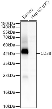
Western blot analysis of various lysates using CD38 Rabbit pAb (CAB13611) at 1:2000 dilution. Secondary antibody: HRP-conjugated Goat anti-Rabbit IgG (H+L) (CABS014) at 1:10000 dilution. Lysates/proteins: 25 μg per lane. Blocking buffer: 3% nonfat dry milk in TBST. Detection: ECL Enhanced Kit (AbGn00021). Negative control (NC): Hep G2. Exposure time: 30s.
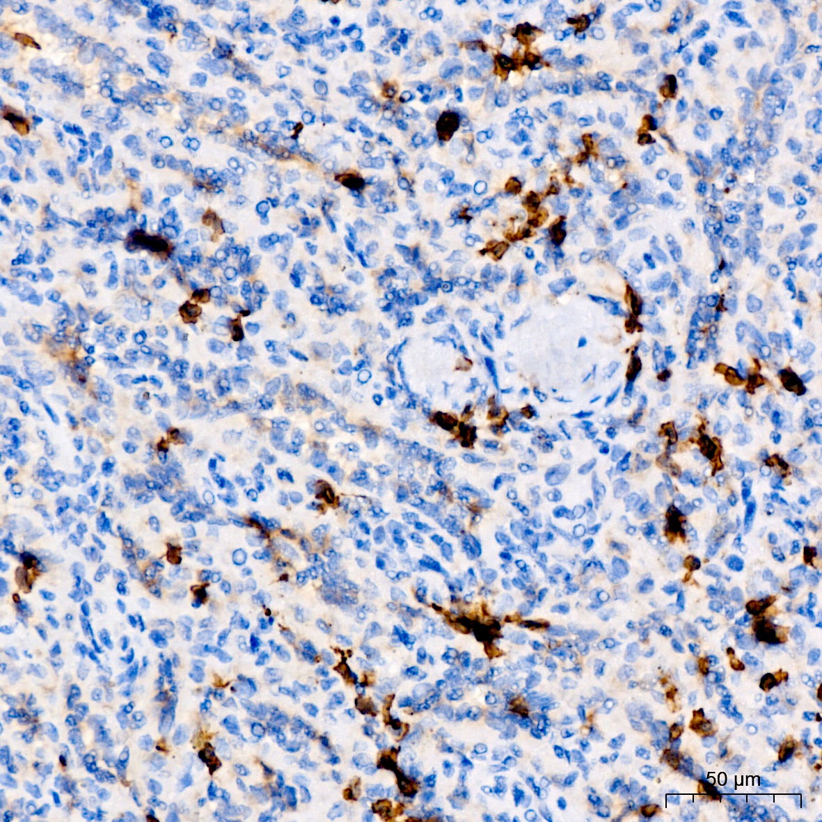
Immunohistochemistry analysis of paraffin-embedded Human spleen tissue using CD38 Rabbit pAb (CAB13611) at a dilution of 1:4000 (40x lens). High pressure antigen retrieval performed with 0.01M Tris-EDTA Buffer (pH 9.0) prior to IHC staining.
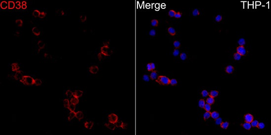
Immunofluorescence analysis of THP-1 cells using CD38 Rabbit pAb (CAB13611) at dilution of 1:100 (40x lens). Secondary antibody: Cy3-conjugated Goat anti-Rabbit IgG (H+L) (CABS007) at 1:500 dilution. Blue: DAPI for nuclear staining.


")
")
")

