Recommended starting concentration is 1 μg/mL. Please optimize the concentration based on your specific assay requirements.
Synonyms:
BHD, FLCL, DENND8B, FLCN
Positive Sample:
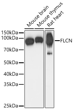
Mouse brain, Mouse thymus, Rat heart,
Cellular Localization:
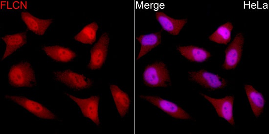
Cytoplasm, Nucleus.
Calculated MW:
64kDa
Observed MW:
70kDa
This gene is located within the Smith-Magenis syndrome region on chromosome 17. Mutations in this gene are associated with Birt-Hogg-Dube syndrome, which is characterized by fibrofolliculomas, renal tumors, lung cysts, and pneumothorax. Alternative splicing of this gene results in two transcript variants encoding different isoforms.
Purification Method
Affinity purification
Gene ID
201163
RRID
AB_2761397
Buffer Information
Store at -20℃. Avoid freeze / thaw cycles. Buffer: PBS with 0.01% thimerosal,50% glycerol,pH7.3.
Western blot analysis of various lysates using FLCN Rabbit pAb (CAB14521) at 1:1000 dilution. Secondary antibody: HRP-conjugated Goat anti-Rabbit IgG (H+L) (CABS014) at 1:10000 dilution. Lysates / proteins: 25 μg per lane. Blocking buffer: 3 % nonfat dry milk in TBST. Detection: ECL Basic Kit (AbGn00020). Exposure time: 1s.
Immunofluorescence analysis of C6 cells using FLCN Rabbit pAb (CAB14521) at dilution of 1:100. Secondary antibody: Cy3-conjugated Goat anti-Rabbit IgG (H+L) (CABS007) at 1:500 dilution. Blue: DAPI for nuclear staining.