The GANAB Antibody (CAB13851) is a high-quality antibody developed for reliable detection and analysis of target proteins. This antibody, produced in rabbits, exhibits high reactivity with human samples and is suitable for use in Western blot applications.The GANAB protein plays a crucial role in maintaining protein homeostasis and proper cell function, making it a target of interest in studies related to protein folding diseases such as neurodegenerative disorders and metabolic syndromes.
This antibody is validated for use in WB, IF/ICC, ELISA applications and has demonstrated reactivity against Human, Mouse, Rat samples.
Product Name:
GANAB Antibody
SKU:
CAB13851
Size:
20μL, 100μL
Reactivity:
Human, Mouse, Rat
Conjugate:
Unconjugated
Immunogen:
Recombinant protein (or fragment).This information is considered to be commercially sensitive.
This gene encodes the alpha subunit of glucosidase II and a member of the glycosyl hydrolase 31 family of proteins. The heterodimeric enzyme glucosidase II plays a role in protein folding and quality control by cleaving glucose residues from immature glycoproteins in the endoplasmic reticulum. Expression of the encoded protein is elevated in lung tumor tissue and in response to UV irradiation. Mutations in this gene cause autosomal-dominant polycystic kidney and liver disease.
Purification Method
Affinity purification
Gene ID
23193
RRID
AB_2760703
Buffer Information
Store at -20℃. Avoid freeze / thaw cycles. Buffer: PBS with 0.01% thimerosal,50% glycerol,pH7.3.
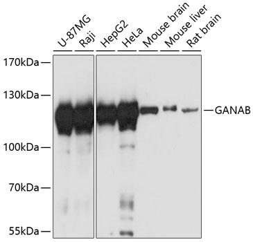
Western blot analysis of various lysates using GANAB Rabbit pAb (CAB13851) at 1:3000 dilution. Secondary antibody: HRP-conjugated Goat anti-Rabbit IgG (H+L) (CABS014) at 1:10000 dilution. Lysates/proteins: 25μg per lane. Blocking buffer: 3% nonfat dry milk in TBST. Detection: ECL Basic Kit (AbGn00020). Exposure time: 10s.
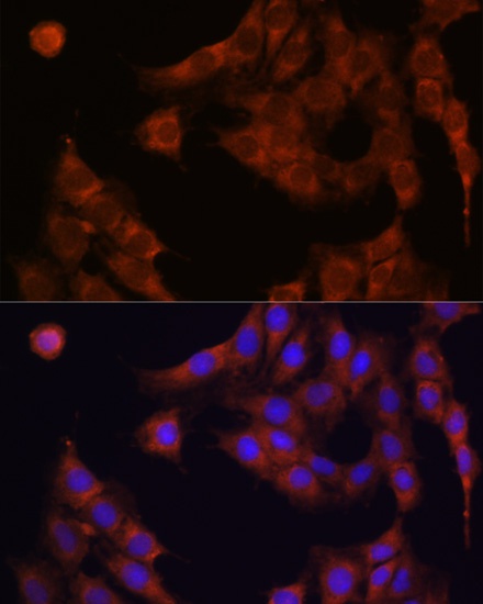
Immunofluorescence analysis of NIH-3T3 cells using GANAB Rabbit pAb (CAB13851) at dilution of 1:100. Secondary antibody: Cy3-conjugated Goat anti-Rabbit IgG (H+L) (CABS007) at 1:500 dilution. Blue: DAPI for nuclear staining.
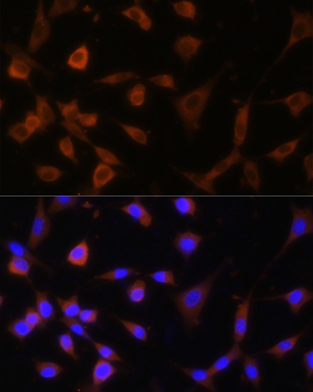
Immunofluorescence analysis of NIH-3T3 cells using GANAB Rabbit pAb (CAB13851) at dilution of 1:100. Secondary antibody: Cy3-conjugated Goat anti-Rabbit IgG (H+L) (CABS007) at 1:500 dilution. Blue: DAPI for nuclear staining.
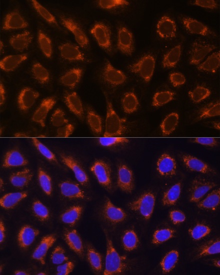
Immunofluorescence analysis of U-2 OS cells using GANAB Rabbit pAb (CAB13851) at dilution of 1:100. Secondary antibody: Cy3-conjugated Goat anti-Rabbit IgG (H+L) (CABS007) at 1:500 dilution. Blue: DAPI for nuclear staining.