The [KO Validated] PDCD4 Antibody (CAB2570) is a high-quality antibody developed for reliable detection and analysis of target proteins. This antibody, produced in rabbits, is highly specific to human samples and has been validated for Western blot applications. By targeting the PDCD4 protein, this antibody allows for accurate detection and analysis in various cell types, making it ideal for studies in molecular biology and cancer research.PDCD4, also known as programmed cell death protein 4, is a tumor suppressor that plays a crucial role in controlling cell cycle progression and apoptosis.
This antibody is validated for use in WB, IHC-P, ELISA applications and has demonstrated reactivity against Human, Mouse samples.
Product Name:
[KO Validated] PDCD4 Antibody
SKU:
CAB2570
Size:
20μL, 100μL
Reactivity:
Human, Mouse
Conjugate:
Unconjugated
Immunogen:
Recombinant protein (or fragment).This information is considered to be commercially sensitive.
Recommended starting concentration is 1 μg/mL. Please optimize the concentration based on your specific assay requirements.
Synonyms:
H731, D4
Positive Sample:
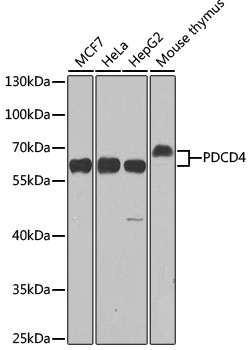
293T, HeLa, MCF7, Mouse thymus
Cellular Localization:
Cytoplasm, Nucleus.
Calculated MW:
52kDa
Observed MW:
60kDa
This gene is a tumor suppressor and encodes a protein that binds to the eukaryotic translation initiation factor 4A1 and inhibits its function by preventing RNA binding. Alternative splicing results in multiple transcript variants.
Purification Method
Affinity purification
Gene ID
27250
RRID
AB_2863014
Buffer Information
Store at -20℃. Avoid freeze / thaw cycles. Buffer: PBS containing 50% glycerol, preserved with proclin300 or sodium azide, pH 7.3.
Western blot analysis of various lysates using [KO Validated] PDCD4 Rabbit pAb (CAB2570) at 1:1000 dilution. Secondary antibody: HRP-conjugated Goat anti-Rabbit IgG (H+L) (CABS014) at 1:10000 dilution. Lysates/proteins: 25μg per lane. Blocking buffer: 3% nonfat dry milk in TBST. Detection: ECL Enhanced Kit (AbGn00021). Exposure time: 90s.
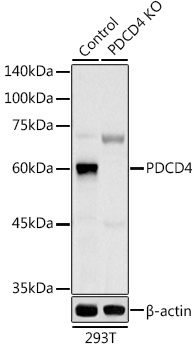
Western blot analysis of lysates from wild type (WT) and PDCD4 knockout (KO) 293T cells, using [KO Validated] PDCD4 Rabbit pAb (CAB2570) at 1:1000 dilution. Secondary antibody: HRP-conjugated Goat anti-Rabbit IgG (H+L) (CABS014) at 1:10000 dilution. Lysates/proteins: 25μg per lane. Blocking buffer: 3% nonfat dry milk in TBST. Detection: ECL Basic Kit (AbGn00020). Exposure time: 180s.
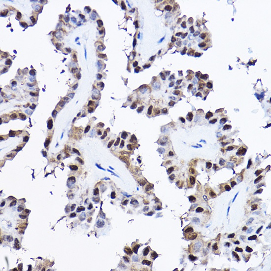
Immunohistochemistry analysis of paraffin-embedded Human thyroid cancer using [KO Validated] PDCD4 Rabbit pAb (CAB2570) at dilution of 1:20 (40x lens). High pressure antigen retrieval performed with 0.01M Citrate buffer (pH 6.0) prior to IHC staining.
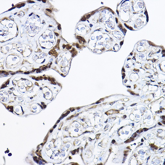
Immunohistochemistry analysis of paraffin-embedded Human placenta using [KO Validated] PDCD4 Rabbit pAb (CAB2570) at dilution of 1:20 (40x lens). High pressure antigen retrieval performed with 0.01M Citrate buffer (pH 6.0) prior to IHC staining.