The MDM2 Monoclonal Antibody (CAB23388) is a high-quality antibody developed for reliable detection and analysis of target proteins. This monoclonal antibody, generated using advanced techniques, is specifically engineered to target MDM2 in human samples, offering high specificity and reliability in experiments.MDM2 is a crucial protein in the cellular pathway that controls the activity of the tumor suppressor protein p53, which plays a pivotal role in preventing the development of cancer. Dysregulation of MDM2 can lead to uncontrolled cell growth and proliferation, characteristic of cancer cells. By targeting MDM2 with this monoclonal antibody, researchers can explore the mechanisms underlying cancer development and potentially identify new therapeutic targets for cancer treatment.
This antibody is validated for use in WB, ELISA applications and has demonstrated reactivity against Human samples.
Product Name:
MDM2 Monoclonal Antibody
SKU:
CAB23388
Size:
20μL, 100μL
Reactivity:
Human
Clone Number:
ARC60296
Conjugate:
Unconjugated
Immunogen:
Synthetic peptide. This information is considered to be commercially sensitive.
This gene encodes a nuclear-localized E3 ubiquitin ligase. The encoded protein can promote tumor formation by targeting tumor suppressor proteins, such as p53, for proteasomal degradation. This gene is itself transcriptionally-regulated by p53. Overexpression or amplification of this locus is detected in a variety of different cancers. There is a pseudogene for this gene on chromosome 2. Alternative splicing results in a multitude of transcript variants, many of which may be expressed only in tumor cells.
Purification Method
Affinity purification
Gene ID
4193
Buffer Information
Store at -20℃. Avoid freeze / thaw cycles. Buffer: PBS containing 50% glycerol and 0.05% BSA, preserved with proclin300 or sodium azide, pH 7.3.
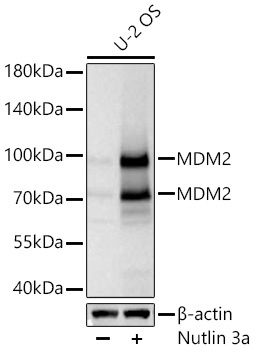
Western blot analysis of lysates from U-2 OS cells using MDM2 Rabbit mAb (CAB23388) at 1:5000 dilution incubated at room temperature for 1.5 hours. U-2 OS cells were treated with Nutlin 3a (10 μM) at 37℃ for 24 hrs Secondary antibody: HRP-conjugated Goat anti-Rabbit IgG (H+L) (CABS014) at 1:10000 dilution. Lysates/proteins: 25 μg per lane. Blocking buffer: 3% nonfat dry milk in TBST. Detection: ECL Basic Kit (AbGn00020). Exposure time: 45s.
 at 1:2000 dilution. U2OS cells were treated by Nutlin 3a(10μM ) at 37℃ for 24 hours. Secondary antibody: HRP Goat Anti-Rabbit IgG (H+L) at 1:10000 dilution. Lysates/proteins: 25μg per lane. Blocking buffer: 3% nonfat dry milk in TBST.")
 at 1:2000 dilution. U2OS cells were treated by Nutlin 3a(10μM ) at 37℃ for 24 hours. Secondary antibody: HRP Goat Anti-Rabbit IgG (H+L) at 1:10000 dilution. Lysates/proteins: 25μg per lane. Blocking buffer: 3% nonfat dry milk in TBST.")

![Anti-MDM2 [BP6203] Monoclonal Antibody (AGMB06453)](https://cdn11.bigcommerce.com/s-h68l9z2lnx/images/stencil/590x590/products/277734/732335/anti-mdm2-bp6203-monoclonal-antibody-agmb06453__75043.1777186523.jpg?c=2 "Anti-MDM2 [BP6203] Monoclonal Antibody (AGMB06453)")
![Anti-Phospho-MDM2 (Ser166) [R08-8C7] Monoclonal Antibody (AGMB05080)](https://cdn11.bigcommerce.com/s-h68l9z2lnx/images/stencil/590x590/products/276365/677165/anti-phospho-mdm2-ser166-r08-8c7-monoclonal-antibody-agmb05080__79709.1773031715.jpg?c=2 "Anti-Phospho-MDM2 (Ser166) [R08-8C7] Monoclonal Antibody (AGMB05080)")

