The OPA1 Antibody (CAB9833) is a high-quality antibody developed for reliable detection and analysis of target proteins. This antibody, produced in rabbits, exhibits high reactivity with human samples and has been validated for use in Western blot applications. By binding specifically to OPA1, this antibody allows for precise detection and analysis of the protein in various cell types, making it a versatile option for studies in mitochondrial biology and related diseases.OPA1, also known as optic atrophy 1 protein, plays a crucial role in maintaining mitochondrial function and integrity.
This antibody is validated for use in WB, IHC-P, IF/ICC, ELISA applications and has demonstrated reactivity against Human, Mouse, Rat samples.
Product Name:
OPA1 Antibody
SKU:
CAB9833
Size:
20μL, 100μL
Reactivity:
Human, Mouse, Rat
Conjugate:
Unconjugated
Immunogen:
Recombinant protein (or fragment).This information is considered to be commercially sensitive.
The protein encoded by this gene is a nuclear-encoded mitochondrial protein with similarity to dynamin-related GTPases. The encoded protein localizes to the inner mitochondrial membrane and helps regulate mitochondrial stability and energy output. This protein also sequesters cytochrome c. Mutations in this gene have been associated with optic atrophy type 1, which is a dominantly inherited optic neuropathy resulting in progressive loss of visual acuity, leading in many cases to legal blindness.
Purification Method
Affinity purification
Gene ID
4976
RRID
AB_2770723
Buffer Information
Store at -20℃. Avoid freeze / thaw cycles. Buffer: PBS containing 50% glycerol, preserved with proclin300 or sodium azide, pH 7.3.
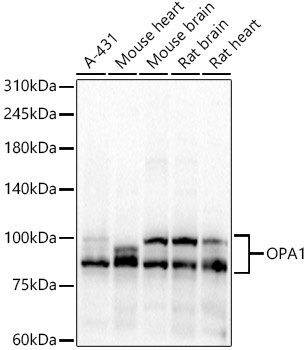
Western blot analysis of various lysates, using OPA1 Rabbit pAb (CAB9833) at 1:7000 dilution. Secondary antibody: HRP-conjugated Goat anti-Rabbit IgG (H+L) (CABS014) at 1:10000 dilution. Lysates/proteins: 25μg per lane. Blocking buffer: 3% nonfat dry milk in TBST. Detection: ECL Basic Kit (AbGn00020). Exposure time: 30s.
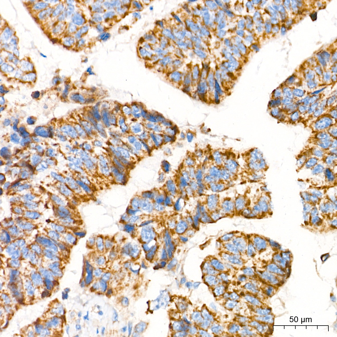
Immunohistochemistry analysis of paraffin-embedded Human colon carcinoma tissue using OPA1 Rabbit pAb (CAB9833) at a dilution of 1:1000 (40x lens). High pressure antigen retrieval performed with 0.01M Tris-EDTA Buffer (pH 9.0) prior to IHC staining.
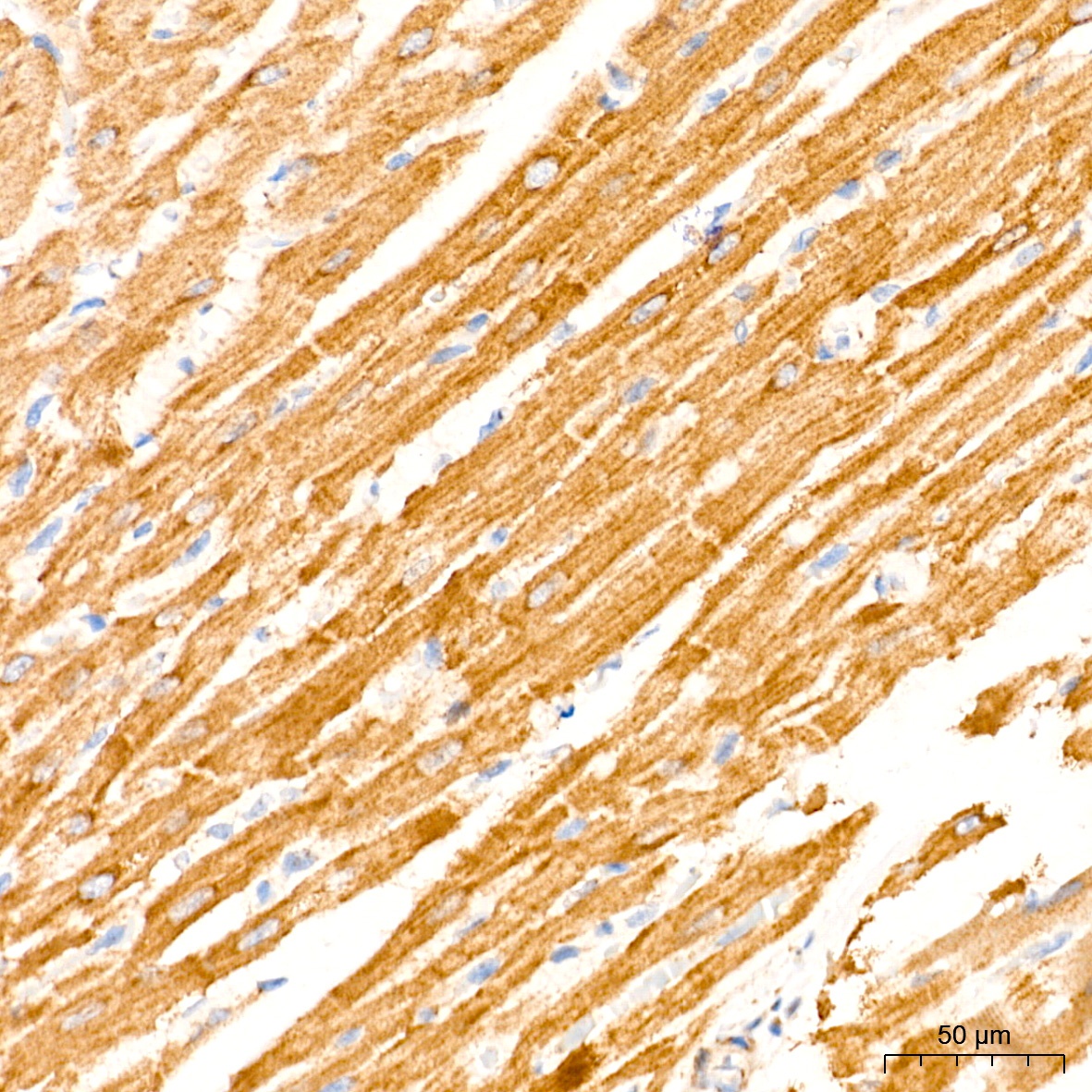
Immunohistochemistry analysis of paraffin-embedded Rat heart tissue using OPA1 Rabbit pAb (CAB9833) at a dilution of 1:1000 (40x lens). High pressure antigen retrieval performed with 0.01M Tris-EDTA Buffer (pH 9.0) prior to IHC staining.