The PDIA6 Monoclonal Antibody (CAB4260) is a high-quality antibody developed for reliable detection and analysis of target proteins. This antibody, produced using rabbit monoclonal technology, provides high specificity and sensitivity in detecting PDIA6 in human samples, making it a reliable choice for Western blot applications.PDIA6, also known as P5, is a multifunctional protein that plays a crucial role in maintaining cellular protein quality control and redox balance. Dysregulation of PDIA6 has been linked to various diseases, including cancer, neurodegenerative disorders, and diabetes, making it a promising target for therapeutic interventions.
This antibody is validated for use in WB, IF/ICC, ELISA applications and has demonstrated reactivity against Human, Mouse, Rat samples.
Product Name:
PDIA6 Monoclonal Antibody
SKU:
CAB4260
Size:
20μL, 100μL
Reactivity:
Human, Mouse, Rat
Clone Number:
ARC0944
Conjugate:
Unconjugated
Immunogen:
Synthetic peptide. This information is considered to be commercially sensitive.
This gene encodes a member of the disulfide isomerase (PDI) family of endoplasmic reticulum (ER) proteins that catalyze protein folding and thiol-disulfide interchange reactions. The encoded protein has an N-terminal ER-signal sequence, two catalytically active thioredoxin (TRX) domains, a TRX-like domain, and a C-terminal ER-retention sequence. This protein inhibits the aggregation of misfolded proteins and exhibits both isomerase and chaperone activity. Alternative splicing results in multiple transcript variants encoding different isoforms.
Purification Method
Affinity purification
Gene ID
10130
RRID
AB_2863220
Buffer Information
Store at -20℃. Avoid freeze / thaw cycles. Buffer: PBS containing 50% glycerol and 0.05% BSA, preserved with proclin300 or sodium azide, pH 7.3.
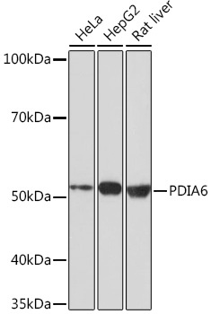
Western blot analysis of various lysates using PDIA6 Rabbit mAb (CAB4260) at 1:1000 dilution. Secondary antibody: HRP-conjugated Goat anti-Rabbit IgG (H+L) (CABS014) at 1:10000 dilution. Lysates/proteins: 25μg per lane. Blocking buffer: 3% nonfat dry milk in TBST. Detection: ECL Basic Kit (AbGn00020). Exposure time: 3min.
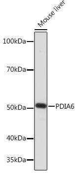
Western blot analysis of lysates from Mouse liver, using PDIA6 Rabbit mAb (CAB4260) at 1:1000 dilution. Secondary antibody: HRP-conjugated Goat anti-Rabbit IgG (H+L) (CABS014) at 1:10000 dilution. Lysates/proteins: 25μg per lane. Blocking buffer: 3% nonfat dry milk in TBST. Detection: ECL Enhanced Kit (AbGn00021). Exposure time: 3min.


![Anti-PDIA6 [R08-6B3] Monoclonal Antibody (AGMB00740)](https://cdn11.bigcommerce.com/s-h68l9z2lnx/images/stencil/590x590/products/272029/695143/anti-pdia6-r08-6b3-monoclonal-antibody-agmb00740__04897.1774514729.jpg?c=2 "Anti-PDIA6 [R08-6B3] Monoclonal Antibody (AGMB00740)")


