The UFD1L Antibody (CAB3255) is a high-quality antibody developed for reliable detection and analysis of target proteins. This antibody, produced in rabbits, exhibits high reactivity with human samples and is validated for use in Western blot applications. It specifically binds to the UFD1L protein, allowing for accurate detection and analysis in a variety of cell types.UFD1L is a crucial component of the ubiquitin-proteasome system, which plays a pivotal role in the degradation of misfolded or damaged proteins. Dysregulation of this system has been implicated in various diseases, including neurodegenerative disorders and cancer.
This antibody is validated for use in WB, IF/ICC, ELISA applications and has demonstrated reactivity against Human, Mouse samples.
Product Name:
UFD1L Antibody
SKU:
CAB3255
Size:
20μL, 100μL
Reactivity:
Human, Mouse
Conjugate:
Unconjugated
Immunogen:
Recombinant protein (or fragment).This information is considered to be commercially sensitive.
Recommended starting concentration is 1 μg/mL. Please optimize the concentration based on your specific assay requirements.
Synonyms:
UFD1L
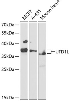
Positive Sample:
MCF7, A-431, Mouse heart
Cellular Localization:
Cytoplasm, Nucleus, Cytosol.
Calculated MW:
35kDa
Observed MW:
37kDa
The protein encoded by this gene forms a complex with two other proteins, nuclear protein localization-4 and valosin-containing protein, and this complex is necessary for the degradation of ubiquitinated proteins. In addition, this complex controls the disassembly of the mitotic spindle and the formation of a closed nuclear envelope after mitosis. Mutations in this gene have been associated with Catch 22 syndrome as well as cardiac and craniofacial defects. Alternative splicing results in multiple transcript variants encoding different isoforms. A related pseudogene has been identified on chromosome 18.
Purification Method
Affinity purification
Gene ID
7353
RRID
AB_2765010
Buffer Information
Store at -20℃. Avoid freeze / thaw cycles. Buffer: PBS containing 50% glycerol, preserved with proclin300 or sodium azide, pH 7.3.
Western blot analysis of various lysates using UFD1L Rabbit pAb (CAB3255) at 1:1000 dilution. Secondary antibody: HRP-conjugated Goat anti-Rabbit IgG (H+L) (CABS014) at 1:10000 dilution. Lysates/proteins: 25μg per lane. Blocking buffer: 3% nonfat dry milk in TBST. Detection: ECL Basic Kit (AbGn00020). Exposure time: 90s.
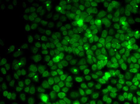
Immunofluorescence analysis of MCF7 cells using UFD1L Rabbit pAb (CAB3255).Secondary antibody: Cy3-conjugated Goat anti-Rabbit IgG (H+L) (CABS007) at 1:500 dilution.


 at 1:10000 dilution. Lysates/proteins: 25ug per lane. Blocking buffer: 3% nonfat dry milk in TBST. Detection: ECL Basic Kit. Exposure time: 30s.")

