HSPA1A Polyclonal Antibody (CAB20819)
- SKU:
- CAB20819
- Product Type:
- Antibody
- Antibody Type:
- Secondary Antibody
- Host Species:
- Goat
- Isotype:
- IgG
. Blue: DAPI for nuclear staining.")
. Blue: DAPI for nuclear staining.")
. Blue: DAPI for nuclear staining.")
. Blue: DAPI for nuclear staining.")
. Blue: DAPI for nuclear staining.")
. Perform high pressure antigen retrieval with 10 mM citrate buffer pH 6. 0 before commencing with IHC staining protocol.")
. Perform high pressure antigen retrieval with 10 mM citrate buffer pH 6. 0 before commencing with IHC staining protocol.")
 at 1:10000 dilution. Lysates/proteins: 25ug per lane. Blocking buffer: 3% nonfat dry milk in TBST. Detection: ECL Basic Kit. Exposure time: 10s.")
 at 1:10000 dilution. Lysates/proteins: 25ug per lane. Blocking buffer: 3% nonfat dry milk in TBST. Detection: ECL Basic Kit. Exposure time: 0. 5s.")
. Blue: DAPI for nuclear staining.")
. Blue: DAPI for nuclear staining.")
. Blue: DAPI for nuclear staining.")
. Blue: DAPI for nuclear staining.")
. Blue: DAPI for nuclear staining.")
. Perform high pressure antigen retrieval with 10 mM citrate buffer pH 6. 0 before commencing with IHC staining protocol.")
. Perform high pressure antigen retrieval with 10 mM citrate buffer pH 6. 0 before commencing with IHC staining protocol.")
 at 1:10000 dilution. Lysates/proteins: 25ug per lane. Blocking buffer: 3% nonfat dry milk in TBST. Detection: ECL Basic Kit. Exposure time: 10s.")
 at 1:10000 dilution. Lysates/proteins: 25ug per lane. Blocking buffer: 3% nonfat dry milk in TBST. Detection: ECL Basic Kit. Exposure time: 0. 5s.")
Frequently bought together:
Description
| Product Name: | HSPA1A Polyclonal Antibody |
| SKU: | CAB20819 |
| Size: | 20uL, 100uL |
| Isotype: | IgG |
| Host Species: | Rabbit |
| Reactivity: | Human,Mouse,Rat |
| Immunogen: | A synthetic peptide corresponding to a sequence within amino acids 500-600 of human HSP70 (NP_005336.3). |
| Sequence: | KITI TNDK GRLS KEEI ERMV QEAE KYKA EDEV QRER VSAK NALE SYAF NMKS AVED EGLK GKIS EADK KKVL DKCQ EVIS WLDA NTLA EKDE FEHK RKEL E |
| Tested Applications: | WB IHC-P IF/ICC ELISA |
| Recommended Dilution: | WB,1:500 - 1:1000 IHC-P,1:50 - 1:200 IF/ICC,1:50 - 1:200 |
| Synonyms: | HEL-S-103; HSP70-1; HSP70-1A; HSP70.1; HSP70I; HSP72; HSPA1; HSP70 |
| Positive Sample: | 293T,HeLa,A-549,NIH/3T3,C6,Mouse brain,Mouse lung,Rat heart,Rat liver |
| Conjugate: | Unconjugated |
| Cellular Localization: | blood microparticle, centriole, centrosome, cytoplasm, cytosol, endoplasmic reticulum, extracellular exosome, extracellular region, focal adhesion, mitochondrion, nuclear speck, nucleoplasm, nucleus, perinuclear region of cytoplasm, plasma membrane |
| Calculated MW: | 70kDa |
| Observed MW: | 70kDa |
This intronless gene encodes a 70kDa heat shock protein which is a member of the heat shock protein 70 family. In conjuction with other heat shock proteins, this protein stabilizes existing proteins against aggregation and mediates the folding of newly translated proteins in the cytosol and in organelles. It is also involved in the ubiquitin-proteasome pathway through interaction with the AU-rich element RNA-binding protein 1. The gene is located in the major histocompatibility complex class III region, in a cluster with two closely related genes which encode similar proteins. [provided by RefSeq, Jul 2008]
| Purification Method: | Affinity purification |
| Gene ID: | 3303/3304 |
| Storage Buffer: | Store at -20℃. Avoid freeze / thaw cycles.Buffer: PBS with 0.05% proclin300,50% glycerol,pH7.3. |
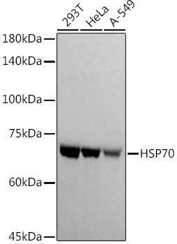 | Western blot analysis of various lysates using HSP70 Rabbit pAb (CAB20819) at 1:1000 dilution.Secondary antibody: HRP Goat Anti-Rabbit IgG (H+L) (CABS014) at 1:10000 dilution.Lysates/proteins: 25μg per lane.Blocking buffer: 3% nonfat dry milk in TBST.Detection: ECL Basic Kit (AbGn00020).Exposure time: 0.5s. |
Related Products


HNRRabbit Polyclonal Antibody Rabbit Polyclonal Antibody (CAB17497)
€139 - €419