
Blood Coagulation Pathway: Factors, Cascade & Disorders [Guide]
Blood Coagulation Pathway: Factors, Cascade and Disorders
The coagulation cascade is usually taught as two pathways converging on a common one. That framework maps neatly onto the laboratory tests but not onto what happens in a blood vessel, where tissue factor initiates and the intrinsic components amplify. This guide covers both models, the factors and complexes involved, the natural anticoagulants that restrain them, and the bleeding and thrombotic disorders that follow when the balance fails.
Browse coagulation assays →Key takeaways
- Coagulation is a cascade of proteolytic activations in which each factor activates the next, amplifying a small initiating signal into a fibrin clot.
- Prothrombin is converted to thrombin by the prothrombinase complex — FXa with FVa, calcium and phospholipid — not by FVIIa.
- The intrinsic and extrinsic pathways converge at factor X activation; the common pathway runs from FXa to fibrin, and is called common because both routes meet there.
- The two-pathway model is an in-vitro construct that maps to aPTT and PT. In vivo, tissue factor initiates and the intrinsic factors amplify — which is why factor XII deficiency causes no bleeding.
- Calcium and phospholipid surfaces are essential cofactors for the tenase and prothrombinase complexes, but contact activation of factor XII does not require calcium.
- Natural anticoagulants — antithrombin, protein C and S, and tissue factor pathway inhibitor — confine clotting to the site of injury.
- Haemophilia A and B are deficiencies of factors VIII and IX; von Willebrand disease is the commonest inherited bleeding disorder.
Contents
- Haemostasis in outline
- The coagulation factors
- Intrinsic (contact activation) pathway
- Extrinsic (tissue factor) pathway
- The common pathway
- Why the cascade model is not the whole story
- Natural anticoagulants and fibrinolysis
- Laboratory testing
- Coagulation disorders
- Choosing coagulation assays
- Frequently asked questions
Coagulation ELISA kits
Clotting-factor activity is measured by functional assays, but the markers below quantify the substrate, the products of activation, the inhibitory complexes and fibrinolysis — which is what most research work actually needs.
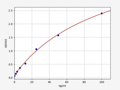
Human Fibrinogen ELISA Kit
Factor I — the soluble precursor thrombin converts into the fibrin clot.
View kit →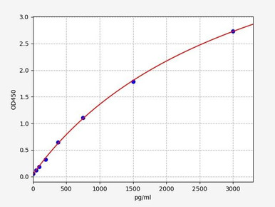
Human Prothrombin Fragment 1+2 ELISA Kit
Released when prothrombin is cleaved, so it reports in vivo thrombin generation directly.
View kit →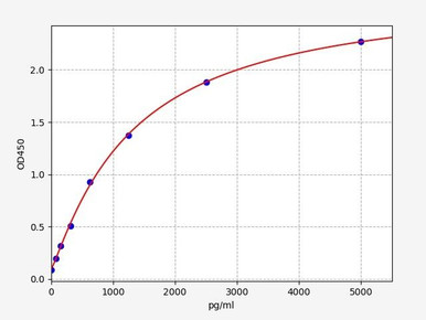
Human Thrombin-Antithrombin Complex ELISA Kit
Quantifies thrombin that has been neutralised by antithrombin — a measure of both generation and inhibition.
View kit →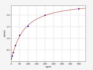
Human Antithrombin-III ELISA Kit
The principal natural inhibitor of thrombin and factor Xa; deficiency causes thrombophilia.
View kit →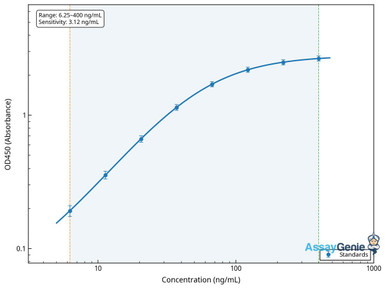
Human D-Dimer ELISA Kit
A fibrin degradation product — evidence that clot has formed and been broken down.
View kit →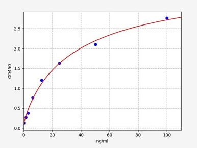
Human von Willebrand Factor ELISA Kit
Mediates platelet adhesion and carries factor VIII; central to the commonest inherited bleeding disorder.
View kit →Haemostasis in outline
Haemostasis stops bleeding without clotting the whole circulation, and it does so in two coordinated stages.
Primary haemostasis is platelet-driven: platelets adhere to exposed collagen and von Willebrand factor at the injury site, activate, change shape and aggregate into a plug. This happens within seconds but the plug is fragile.
Secondary haemostasis is the coagulation cascade, which generates thrombin and converts fibrinogen into a fibrin mesh that stabilises the platelet plug into a durable clot. The two are interdependent: activated platelets provide the phospholipid surface the cascade requires, and thrombin is among the strongest platelet activators.
A clarification worth making early: platelets are cells, not clotting factors, and calcium is a cofactor rather than a factor in the enzymatic sense, although it was historically designated factor IV. The numbered coagulation factors are plasma proteins, mostly synthesised in the liver, and mostly circulating as inactive zymogens until proteolytically cleaved. Clotting is not controlled by regulating their release — they are present continuously — but by controlling their activation.
The coagulation factors
Thirteen numbers were assigned historically, though factor VI was retired once it was recognised as activated factor V rather than a distinct protein.
| Factor | Name | Role |
|---|---|---|
| I | Fibrinogen | Soluble precursor of fibrin; the clot substrate |
| II | Prothrombin | Precursor of thrombin, the central enzyme |
| III | Tissue factor | Membrane receptor exposed on injury; initiates coagulation in vivo |
| IV | Calcium ions | Cofactor bridging factors to phospholipid surfaces |
| V | Proaccelerin | Cofactor for factor Xa in the prothrombinase complex |
| VII | Proconvertin | Partners tissue factor to activate factor X |
| VIII | Antihaemophilic factor | Cofactor for factor IXa; deficient in haemophilia A |
| IX | Christmas factor | Activates factor X with factor VIIIa; deficient in haemophilia B |
| X | Stuart-Prower factor | Convergence point of both pathways; forms prothrombinase |
| XI | Thromboplastin antecedent | Activates factor IX |
| XII | Hageman factor | Contact activation; deficiency causes no bleeding |
| XIII | Fibrin-stabilising factor | Cross-links fibrin to consolidate the clot |
Factors II, VII, IX and X require vitamin K for post-translational gamma-carboxylation, which is what lets them bind calcium and anchor to phospholipid. This is the basis of warfarin’s action and of the vitamin K dependence of the prothrombin time.
Intrinsic (contact activation) pathway
The intrinsic pathway is triggered when blood contacts a negatively charged surface — exposed subendothelial collagen in vivo, or glass and kaolin in the laboratory. All its components are already present in plasma, which is where the name comes from.
- Factor XII (Hageman factor) autoactivates on the charged surface to factor XIIa, assisted by prekallikrein and high molecular weight kininogen. HMWK acts here, at contact activation — not further down the pathway.
- Factor XI is cleaved by factor XIIa to factor XIa. Thrombin can also activate factor XI, which is one of the feedback loops that amplifies the response.
- Factor IX is activated by factor XIa to factor IXa, in a calcium-dependent step.
- The intrinsic tenase complex assembles: factor IXa with its cofactor factor VIIIa, calcium, and a phospholipid surface supplied by activated platelets. Note that factor VIII must itself be activated by thrombin before it can serve as cofactor.
- Factor X is activated by that complex to factor Xa, which enters the common pathway.
The intrinsic tenase complex is roughly fifty-fold more efficient at activating factor X than the extrinsic complex, which is why this arm dominates once coagulation is under way even though it does not start it.
Extrinsic (tissue factor) pathway
The extrinsic pathway is triggered by tissue damage — that is precisely its defining feature. Tissue factor is a transmembrane protein expressed on subendothelial cells, fibroblasts and smooth muscle, normally separated from blood by intact endothelium. Vessel injury exposes it, and coagulation begins. It is called extrinsic because tissue factor comes from outside the blood, not because the trigger is something other than injury.
- Tissue factor (factor III) is exposed at the site of injury and binds circulating factor VII.
- The TF-FVIIa complex forms as bound factor VII is rapidly activated. This is the extrinsic tenase.
- Factor X is activated to factor Xa by that complex, entering the common pathway. The complex also activates factor IX, which recruits the intrinsic arm — the crosstalk that makes the two-pathway separation artificial.
This route is fast, producing an initial burst of thrombin within seconds, but it is quickly shut down by tissue factor pathway inhibitor. That initial thrombin is not enough to form a clot on its own; its main job is to activate factors V, VIII and XI and platelets, so that the far more productive intrinsic tenase can take over.
The common pathway
The common pathway is the segment both routes share, running from factor X activation to a cross-linked fibrin clot. It is called common because the intrinsic and extrinsic pathways converge on it — not because it contains all the clotting factors.
- Prothrombinase assembles. Factor Xa combines with cofactor factor Va, calcium and a phospholipid surface. This complex — also called prothrombinase or the prothrombin activator complex — is what cleaves prothrombin.
- Thrombin is generated. Prothrombin (factor II) is cleaved to thrombin (factor IIa) by the prothrombinase complex. It is not converted by factor VIIa; FVIIa acts upstream, on factors X and IX. Cleavage also releases prothrombin fragment 1+2, which is why that fragment is a direct marker of thrombin generation.
- Fibrin forms. Thrombin cleaves fibrinogen to fibrin monomers, which polymerise into a mesh.
- The clot is stabilised. Thrombin activates factor XIII, which covalently cross-links fibrin strands, making the clot mechanically strong and resistant to premature lysis.
- Thrombin amplifies its own production by activating factors V, VIII and XI and by activating platelets — a positive feedback loop that converts a trickle into a burst.
On calcium: it is required for the assembly of both tenase complexes and prothrombinase, because gamma-carboxylated factors bind phospholipid through calcium bridges. Contact activation of factor XII, however, is not calcium-dependent — which is why citrated plasma can still undergo contact activation in a tube.
Why the cascade model is not the whole story
The two-pathway cascade was proposed in the 1960s and remains the standard teaching model, largely because it maps so cleanly onto the two screening tests. It is nonetheless a description of what happens in a test tube rather than in a person.
The clearest evidence is clinical. Factor XII deficiency causes no bleeding disorder at all, despite factor XII sitting at the top of the intrinsic pathway. If the cascade model described reality, losing the initiator would be catastrophic. Equally, haemophilia A and B — deficiencies of the intrinsic factors VIII and IX — cause severe bleeding even though the extrinsic pathway is intact and should, on the cascade model, compensate.
The modern cell-based model resolves this in three overlapping phases. Initiation: tissue factor exposure generates a small amount of thrombin. Amplification: that thrombin activates platelets and factors V, VIII and XI, priming the system. Propagation: intrinsic tenase assembled on activated platelet surfaces generates the large thrombin burst that actually produces a clot. Coagulation is therefore one integrated process localised to a cell surface, with the intrinsic factors serving as the amplifier rather than an independent pathway.
The cascade model remains worth knowing because the laboratory tests are built on it. But it explains the tests, not the biology.
Natural anticoagulants and fibrinolysis
A cascade with this much positive feedback needs restraining, or a local injury would clot the entire circulation. Four mechanisms confine it.
- Antithrombin irreversibly inhibits thrombin and factor Xa, and to a lesser extent factors IXa, XIa and XIIa. Heparin accelerates it several thousand-fold, which is how heparin works. Antithrombin deficiency is a recognised inherited thrombophilia.
- Protein C and protein S. Thrombin bound to endothelial thrombomodulin changes specificity and activates protein C, which with protein S inactivates factors Va and VIIIa — an elegant switch by which thrombin turns off its own amplifiers. Factor V Leiden resists this inactivation and is the commonest inherited thrombophilia.
- Tissue factor pathway inhibitor shuts down the TF-FVIIa complex shortly after initiation, which is why the extrinsic burst is brief and the intrinsic amplifier is needed.
- Endothelial surface. Intact endothelium is actively antithrombotic, expressing thrombomodulin and heparan sulphate and releasing prostacyclin and nitric oxide to inhibit platelets.
Fibrinolysis
Clots are eventually removed. Tissue plasminogen activator converts plasminogen to plasmin, which digests fibrin into soluble fragments. D-dimer is one of those fragments, and because it only exists if fibrin was formed and then lysed, it is a direct marker of coagulation activity followed by breakdown. Raised D-dimer is sensitive but not specific — it rises in infection, malignancy, pregnancy and after surgery — which is why it is used mainly to exclude thrombosis rather than confirm it.
Laboratory testing
The two screening tests correspond directly to the cascade model, which is the model’s main practical value.
| Test | Pathway assessed | Prolonged by |
|---|---|---|
| Prothrombin time (PT / INR) | Extrinsic and common — factors VII, X, V, II, I | Warfarin, vitamin K deficiency, liver disease, factor VII deficiency |
| Activated partial thromboplastin time (aPTT) | Intrinsic and common — factors XII, XI, IX, VIII, X, V, II, I | Heparin, haemophilia A and B, lupus anticoagulant, factor XII deficiency |
| Thrombin time | Fibrinogen to fibrin conversion only | Heparin, dysfibrinogenaemia, direct thrombin inhibitors |
| Fibrinogen (Clauss) | Functional fibrinogen concentration | Consumption in DIC, liver failure, dilution |
| D-dimer | Fibrin formation and lysis | Thrombosis, but also infection, surgery, pregnancy, malignancy |
Two practical points. Coagulation tests require citrated plasma at a fixed anticoagulant-to-blood ratio, so an underfilled tube gives falsely prolonged times — serum is useless for these assays because the factors have already been consumed. And a prolonged aPTT with a normal PT points to the intrinsic factors, while the reverse points to factor VII; both prolonged suggests the common pathway, liver disease or DIC.
Coagulation disorders
Haemophilia
Haemophilia A results from deficiency of factor VIII and haemophilia B, also called Christmas disease, from deficiency of factor IX. Both are X-linked, present with joint and muscle bleeding rather than the mucosal bleeding typical of platelet disorders, and prolong the aPTT while leaving the PT normal. Severity tracks residual factor activity.
Von Willebrand disease
The commonest inherited bleeding disorder, caused by deficiency or dysfunction of von Willebrand factor. Because vWF both mediates platelet adhesion and carries factor VIII, the condition produces a mixed picture — mucocutaneous bleeding from impaired platelet adhesion, plus a variable reduction in factor VIII.
Thrombocytopenia
A low platelet count, from reduced production, increased destruction or sequestration. Causes include immune thrombocytopenia, drugs, viral infection, marrow disorders and splenomegaly. Bleeding is typically mucocutaneous — petechiae, purpura, epistaxis — rather than deep.
Antiphospholipid syndrome
An autoimmune disorder in which antibodies against phospholipid-binding proteins promote thrombosis and pregnancy morbidity. It carries a characteristic paradox: the lupus anticoagulant prolongs the aPTT in vitro while the patient is prothrombotic in vivo.
Hypercoagulable states
Inherited or acquired conditions tipping the balance toward thrombosis. Inherited causes include factor V Leiden, prothrombin G20210A, and deficiencies of antithrombin, protein C or protein S. Acquired causes include malignancy, immobility, surgery, pregnancy, oestrogen therapy and obesity. Clinical consequences include:
- Deep vein thrombosis — clot in a deep vein, usually of the leg, causing pain, swelling and warmth.
- Pulmonary embolism — embolised clot obstructing pulmonary arteries, causing breathlessness and chest pain. A medical emergency.
- Disseminated intravascular coagulation — widespread activation consuming factors and platelets, so bleeding and thrombosis occur together. Always secondary to another condition such as sepsis, trauma or obstetric complication.
Suspected thrombosis or abnormal bleeding needs clinical assessment. This article describes the biology and is not medical advice.
Choosing coagulation assays
ELISA kits for fibrinogen, prothrombin fragment 1+2, thrombin-antithrombin complex, antithrombin III, D-dimer and von Willebrand factor — for quantifying activation, inhibition and fibrinolysis.
Browse coagulation assays →Frequently asked questions
What converts prothrombin into thrombin?
The prothrombinase complex — factor Xa together with cofactor factor Va, calcium and a phospholipid surface. It is not factor VIIa, which acts upstream on factors X and IX as part of the tissue factor complex.
Why is it called the common pathway?
Because the intrinsic and extrinsic pathways converge on it at factor X activation. It runs from factor Xa through thrombin generation to cross-linked fibrin. It is not called common because it contains all the clotting factors.
What is the difference between the intrinsic and extrinsic pathways?
The extrinsic pathway is initiated by tissue factor exposed on injury — tissue factor comes from outside the blood, hence extrinsic. The intrinsic pathway uses components already present in plasma and is triggered by contact with a negatively charged surface. Both converge at factor X.
If factor XII starts the intrinsic pathway, why does its deficiency cause no bleeding?
Because the cascade model is an in-vitro description. In vivo, coagulation is initiated by tissue factor, and the intrinsic factors act as an amplifier downstream. Factor XII is dispensable for haemostasis, though it does prolong the aPTT.
Is calcium needed for every step?
No. Calcium is essential for assembly of the tenase and prothrombinase complexes, because gamma-carboxylated factors bind phospholipid through calcium bridges. Contact activation of factor XII does not require calcium.
Which tests assess which pathway?
Prothrombin time assesses the extrinsic and common pathways and is prolonged by warfarin. Activated partial thromboplastin time assesses the intrinsic and common pathways and is prolonged by heparin and by haemophilia. Both prolonged suggests the common pathway, liver disease or DIC.
Why must coagulation samples be citrated plasma?
Because citrate chelates calcium reversibly, halting clotting until it is recalcified in the assay. Serum cannot be used at all, since the factors have already been consumed in forming the clot. The anticoagulant-to-blood ratio is fixed, so underfilled tubes give falsely prolonged results.
Rithika Suresh completed her undergraduate degree in Biotechnology in Anna University before completing her masters in Biotechnology at University College Dublin.
Recent Posts
-
Suspected Plague Death at a Siberian Anti-Plague Institute: What Researchers Should Know
Infectious Disease · News AnalysisSuspected Plague Death at a Siberian Anti-Plague Institute: What …6th Oct 2026 -
LPS ELISA Troubleshooting: The Importance of Sample Nature
Quick answer A researcher measuring circulating lipopolysaccharide (LPS) in human serum …25th Sep 2026 -

Sekundärantikörper wählen: Die Regel, die im Labor wirklich zählt
Sekundärantikörper wählen: Die Regel, die im Labor wirklich zählt Wählen Sie einen Sekundärantikörp …24th Sep 2026