
The Strategic Role of Secondary Antibody Incubation Times in Immunodetection Techniques
Secondary Antibody Incubation: Times, Conditions and Troubleshooting
The secondary antibody step determines how much signal you get and how much background comes with it. Incubation time is one lever among several, and rarely the most important — concentration, blocking and washing usually matter more. This guide gives typical times for each technique, explains what actually drives signal-to-noise, and covers the controls that tell you whether the secondary is behaving.
Browse secondary antibodies →Key takeaways
- Typical secondary incubations: 1 hour at room temperature for western blot, 30–60 minutes for ELISA and for IHC. These are starting points, not fixed rules.
- Longer is not better. Specific binding saturates while non-specific binding keeps accumulating, so extending incubation past the plateau costs signal-to-noise rather than gaining signal.
- Titrating the secondary usually improves a blot more than changing its incubation time. Concentration and time are not interchangeable in their effect on background.
- The secondary must be raised against the host species of the primary, and should not cross-react with the sample species — cross-adsorbed secondaries exist for exactly this.
- Run a no-primary control. If bands or staining appear without the primary, the problem is the secondary or the block, and no amount of incubation tuning will fix it.
- Fluorescent conjugates photobleach, so incubate and store them protected from light; enzymatic conjugates do not have that constraint but do have substrate-dependent development.
- Washing between steps often does more for background than incubation time does.
Secondary antibodies and detection reagents
Conjugates for the two commonest primary host species, a fluorescent option, and the dilution, detection and stripping reagents that surround the step.
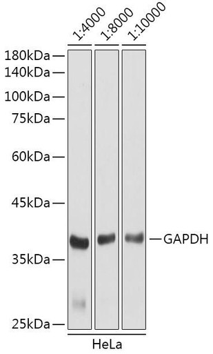
HRP-conjugated Goat anti-Rabbit IgG (H+L)
H+L recognises heavy and light chains — the general-purpose choice for rabbit primaries.
View product →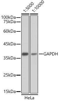
HRP-conjugated Goat anti-Mouse IgG (H+L)
The equivalent for mouse primaries; pair with an ECL substrate matched to your expected abundance.
View product →
GenieFluor 488 Goat Anti-Mouse IgG (H+L)
Fluorescent detection for multiplexing and quantitative work — keep protected from light throughout.
View product →
Quick Block Western Blotting Antibody Dilution Buffer
Diluting the secondary in a blocking-compatible buffer is a direct lever on background.
View product →
Super Sensitivity Chemiluminescent Substrate (ECL)
Substrate sensitivity and secondary concentration trade off — a more sensitive substrate permits a more dilute secondary.
View product →
Western Blot Stripping Buffer
Allows reprobing when a secondary has produced unusable background, without repeating the transfer.
View product →What the secondary antibody does
In indirect detection, the primary antibody binds the target antigen and carries no label. A labelled secondary antibody, raised against immunoglobulin from the primary’s host species, then binds the primary and supplies the detectable signal.
Two consequences follow. First, amplification: a polyclonal secondary recognises several epitopes on each primary molecule, so more than one label is delivered per bound primary. Second, flexibility: one labelled secondary serves every primary raised in that species, which is why laboratories stock few secondaries and many primaries.
Typical incubation times by technique
The original version of this article discussed incubation times without giving any. These are the conventional starting points.
| Technique | Typical time | Temperature | Notes |
|---|---|---|---|
| Western blot | 1 hour | Room temperature | The standard; 30 min often suffices with an abundant target and a sensitive substrate |
| ELISA | 30–60 minutes | Room temperature or 37 °C | Plate assays are diffusion-limited over a short path, so shorter times work well |
| Immunohistochemistry | 30–60 minutes | Room temperature | Polymer detection systems are often shorter still, around 20–30 min |
| Immunocytochemistry | 1 hour | Room temperature | Protect from light for fluorescent conjugates |
| Flow cytometry | 20–30 minutes | On ice, dark | Cold limits internalisation and capping of surface complexes |
A note on a claim in the earlier version: western blot secondaries do not generally need shorter incubation than IHC. In practice the two are broadly comparable, and IHC with a polymer system is frequently the quicker of the two. What differs is the matrix — antibody must penetrate a tissue section, whereas on a membrane the target is surface-accessible.
Why longer is not better
Binding follows saturation kinetics. Specific, high-affinity binding occupies available sites quickly and then plateaus. Non-specific binding — low-affinity, driven by concentration and surface chemistry — does not plateau in the same way and continues to accumulate.
The practical consequence: past the plateau, extra time adds background but not signal. An overnight secondary incubation rarely improves a weak blot; it usually produces the same specific bands on a grey membrane.
If signal is genuinely too low, the productive changes are more primary antibody, a more sensitive substrate, more loaded protein, or a more efficient transfer — not a longer secondary step.
Factors that change the optimum
| Factor | Effect | Adjustment |
|---|---|---|
| Secondary concentration | The dominant driver of both signal and background | Titrate; typical western dilutions run 1:5,000 to 1:20,000 |
| Temperature | Higher temperature speeds binding but also raises non-specific interaction | Room temperature for most work; cold for surface staining |
| Target abundance | Low-abundance targets need more total signal, not more time | Increase primary, use a more sensitive substrate |
| Blocking | Inadequate blocking shows up as background regardless of timing | Match blocker to system; avoid milk with phospho-specific antibodies |
| Washing | Insufficient washing is a very common background source | Three to five washes with detergent-containing buffer between steps |
| Conjugate type | HRP develops fast; fluorophores are read directly and photobleach | Protect fluorescent conjugates from light at every stage |
Milk is worth singling out: it contains casein, a phosphoprotein, so it competes with phospho-specific antibodies and causes weak or absent signal. BSA is the conventional alternative there.
Choosing the right secondary
- Match the host species of the primary. A rabbit primary needs an anti-rabbit secondary. This is the first thing to check when a blot is completely blank.
- Check cross-reactivity with the sample species. Detecting mouse protein with a mouse primary means the anti-mouse secondary will also bind endogenous mouse immunoglobulin. Cross-adsorbed secondaries are pre-depleted against other species to prevent this.
- H+L versus heavy-chain specific. H+L conjugates recognise heavy and light chains and suit general use. Heavy-chain-specific secondaries avoid detecting the ~25 kDa light chain, which matters when blotting immunoprecipitates.
- Match the conjugate to the readout. HRP with ECL for sensitivity, alkaline phosphatase for prolonged colorimetric development, fluorophores for multiplexing and quantitation.
- For multiplex fluorescence, choose spectrally separated fluorophores and cross-adsorbed secondaries, or the channels will bleed into one another.
Optimising: what to change first
Change one variable at a time, in roughly this order of impact:
- Titrate the secondary. A dilution series scored on band intensity against membrane background is the single most informative experiment, and usually reduces reagent use as well.
- Improve washing. More washes, longer washes, adequate volume. Cheap and frequently sufficient.
- Revisit blocking. Blocker type and concentration, and whether the secondary is diluted in blocker.
- Then adjust incubation time, within the ranges above.
- Finally consider the substrate, matching its sensitivity to target abundance rather than defaulting to the most sensitive available.
Controls
| Control | What it shows |
|---|---|
| No-primary (secondary only) | Whether background originates from the secondary or the block — the single most useful control for this step |
| No-secondary | Endogenous enzyme activity or sample autofluorescence |
| Isotype control | Non-specific binding attributable to the primary’s isotype rather than its specificity |
| Positive control lysate or tissue | Confirms the detection chain works when a sample is negative |
| Loading control | Normalises for protein loaded and transfer efficiency |
The no-primary control deserves emphasis. If signal appears without any primary present, adjusting incubation times is wasted effort — the secondary is binding something directly, and the fix is a cross-adsorbed secondary, better blocking, or a more dilute conjugate.
Troubleshooting
| Problem | Likely cause | Action |
|---|---|---|
| No signal at all | Species mismatch between primary and secondary | Confirm the secondary targets the primary’s host species |
| High uniform background | Secondary too concentrated, or inadequate washing | Dilute further; increase wash number and duration |
| Speckled or blotchy background | Aggregated secondary, or dried membrane | Centrifuge the diluted conjugate before use; keep the membrane wet throughout |
| Extra band near 25 or 50 kDa | Light and heavy chains of a precipitating antibody | Use a heavy-chain-specific or conformation-specific secondary |
| Weak phospho-specific signal | Casein in milk competing with the antibody | Block and dilute in BSA instead |
| Fluorescence fading between imaging runs | Photobleaching | Protect from light at every stage; image promptly |
References
- Yannakou, L., Diamandis, E. P., & Souvatzoglou, A. (1987). Effect of incubation time and temperature on the interference of digoxin-like immunoreactive substances in digoxin immunoassays. Therapeutic drug monitoring, 9(4), 461-463.
- Jiang, W., Liu, X., Wu, D., Wang, H., Wang, Y., Chen, H., & Yuan, L. (2015). A simple, rapid one‐step ELISA using antibody–antibody complex. Biotechnology and Applied Biochemistry, 62(1), 126-131.
- Simons, B., Kaplan, H., & Hefford, M. A. (2006). Novel cross-linked enzyme–antibody conjugates for Western blot and ELISA. Journal of immunological methods, 315(1-2), 88-98.
- Kim, S. W., Roh, J., & Park, C. S. (2016). Immunohistochemistry for pathologists: protocols, pitfalls, and tips. Journal of pathology and translational medicine, 50(6), 411.
- Wujcik, E. K., Wei, H., Zhang, X., Guo, J., Yan, X., Sutrave, N., ... & Guo, Z. (2014). Antibody nanosensors: a detailed review. Rsc Advances, 4(82), 43725-43745.
Choosing reagents
HRP and fluorescent secondary antibodies against rabbit and mouse primaries, plus dilution buffer, ECL substrate and stripping buffer.
Browse secondary antibodies →Frequently asked questions
How long should I incubate a secondary antibody?
One hour at room temperature is the standard for western blotting, and 30 to 60 minutes for ELISA and IHC. Flow cytometry surface staining is typically 20 to 30 minutes on ice in the dark. Treat these as starting points and titrate from there.
Will a longer incubation give me more signal?
Rarely. Specific binding saturates and plateaus, while non-specific binding keeps accumulating, so extra time past the plateau adds background rather than signal. If signal is genuinely weak, increase the primary or use a more sensitive substrate.
Should I incubate the secondary overnight at 4 degrees?
It is not usually necessary. Overnight incubation is common for primary antibodies, where it can improve specificity at low concentration. For secondaries the binding is fast and high-affinity, so an hour at room temperature is generally sufficient.
What is the most useful control for this step?
The no-primary control. If you see bands or staining with the secondary alone, the background comes from the secondary or the blocking step, and adjusting incubation time will not fix it.
What does H+L mean on a secondary antibody?
It recognises both the heavy and light chains of immunoglobulin — the general-purpose format. A heavy-chain-specific secondary avoids detecting the light chain around 25 kDa, which is useful when blotting immunoprecipitated samples.
Why is my phospho-specific signal weak?
Frequently because of milk. Casein is a phosphoprotein and competes with phospho-specific antibodies, so blocking and diluting in BSA rather than milk usually restores the signal.
What should I change first when optimising?
Titrate the secondary. Concentration affects signal and background more than incubation time does. After that, improve washing, revisit blocking, and only then adjust the incubation itself.
Tehreem Ali completed her MS in Bioinformatics and conducted her research work at the IOMM lab at GCUF, Pakistan.
Recent Posts
-
LPS ELISA Troubleshooting: The Importance of Sample Nature
Quick answer A researcher measuring circulating lipopolysaccharide (LPS) in human serum …25th Sep 2026 -

Sekundärantikörper wählen: Die Regel, die im Labor wirklich zählt
Sekundärantikörper wählen: Die Regel, die im Labor wirklich zählt Wählen Sie einen Sekundärantikörp …24th Sep 2026 -

Anticuerpos para IHC: cómo validarlos y evitar falsos positivos
Anticuerpos para IHC: cómo validarlos y evitar falsos positivos Use anticuerpos con evidencia publi …24th Sep 2026